3343
Field-map correction in read-out segmented echo planar imaging for reduced spatial distortion in prostate DWI – a phantom study1Physics & Astronomy, University of Manitoba, Winnipeg, MB, Canada, 2Medical Physics, CancerCare Manitoba, Winnipeg, MB, Canada
Synopsis
Diffusion-weighted magnetic resonance imaging (DWI) is routinely used in prostate cancer assessment, but suffers from image distortions primarily due to the tissue-air interface at the rectal cavity. Readout-segmented echo planar imaging (RESOLVE) improves image quality through segmented acquisition of k-space, increasing bandwidth in the phase direction. However, distortions of several millimeters may still exist in RESOLVE images. This study quantified distortions in a prostate phantom by varying the number of RESOLVE segments and using field mapping correction techniques. Field mapping correction decreased image distortion by 28% compared to the 7-segment RESOLVE scan.
Purpose
To assess the use of field map corrections in conventional diffusion-weighted echo planar imaging (EPI), and RESOLVE, using a DWI phantom.Method
The prostate and rectal anatomy was simulated by inserting a cylindrical air cavity with a 1-inch diameter into a container filled with water. A spherical container with a 38mm diameter was filled with water to simulate the prostate. By increasing the angle of the cylindrical cavity with respect to the main field, the inhomogeneity could be increased and image distortion could be controlled. Scans were performed on a 3T Siemens Verio MRI scanner. T2 images were acquired at a FOV of 200x200 mm, slice thickness of 3mm, with a bandwidth of 256 Hz/px and TE/TR = 96/5200ms. Axial DW-EPI images were acquired at a FOV of 220x187mm, slice thickness of 3.6mm, resolution of 160x120 with a phase bandwidth of 11 Hz/px and 6 averages. RESOLVE images were acquired at the same settings with 1 average, with the number of segments ranging from 7-21. Apparent diffusion coefficient (ADC) maps were calculated using b values of 0, 500 and 1000 mm2/s. Field mapping was calculated from gradient echo (GRE) images acquired at echo times of 3.1 and 4.1ms using the same geometry and 4 averages. Field mapping corrections followed the procedure outlined by Jezzard1. Distortions were assessed by measuring the smallest diameter of the spherical volume in the phase encoding direction. Errors in distance measurements were assumed to be 0.7mm, which corresponds to half a pixel.Results
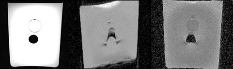
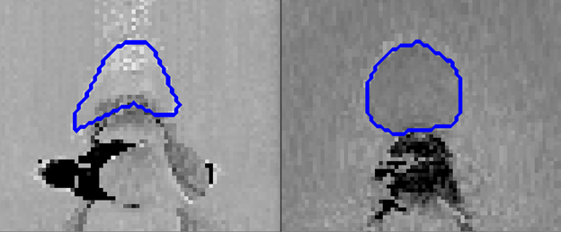
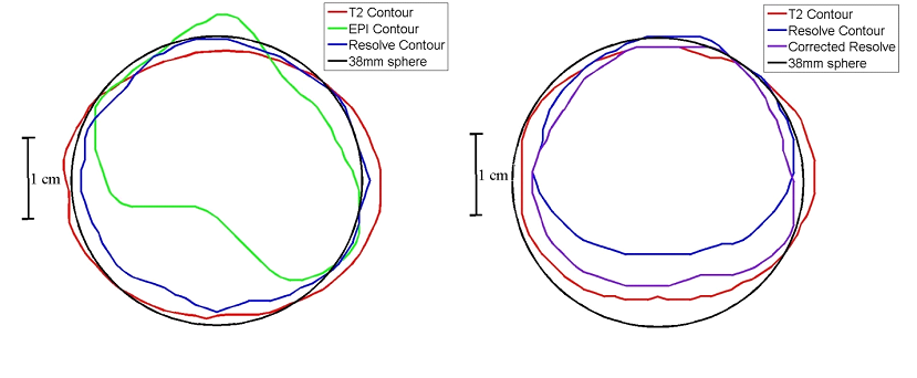
Images of the T2, EPI and RESOLVE images with the air cavity angled 20 degrees to the main field are shown in Figure 1. The RESOLVE image remains undistorted at 7 segments while the EPI image suffers from large distortions. Images acquired with the air cavity angled 45 degrees to the main field are shown in Figure 2. Following the field map correction approach outlined in the previous section, a corrected EPI and corrected RESOLVE image were generated, as shown in Figure 3. The prostate was contoured manually in each image and these contours were overlaid on a perfect 38mm circle at cavity angles of 20 and 45 degrees relative to the main field (Figure 4). The 7-segment RESOLVE contour was distorted by 9.6mm and was reduced to 6.9mm with field mapping, decreasing distortion by 28%. Increasing the number of segments to reduce spatial distortion was also investigated. 13-segment RESOLVE scans reduced distortions by only 7% compared to 7-segment RESOLVE scans. The associated increase in scan time (5:34 min) was similar to that of the GRE acquisitions (6:04 min). The same experiment was carried out for 21 segments, but no additional benefit was seen and distortion reduction remained at 7%.Discussion
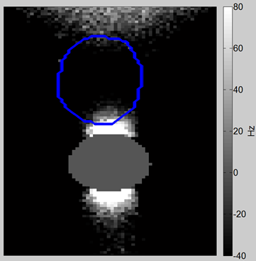
Diffusion MRI is routinely used for assessing tumour aggressiveness in prostate cancer2-4. It is known that DW-EPI is susceptible to image distortion due to its low bandwidth in the phase encoding direction. This complicates its use in a MR-guided radiotherapy treatment setting, since high geometric accuracy is required. Prostate DWI is particularly challenging since the tissue-air interface at the rectal wall introduces local field inhomogeneities5. As shown in Figure 3 and 4, RESOLVE greatly reduces distortions compared to traditional DWI through a segmented acquisition of k-space6, but still suffers from residual distortion in some cases. Our phantom simulates the rectum/prostate anatomy using a cylindrical air cavity to represent rectal gas and a sphere to represent the prostate. The simulated prostate was placed 7.5mm away from the rectal wall. For an infinite cylinder of air with its long axis parallel to the main field, the field strength in water just outside the cavity remains unchanged. Rotating the cavity out of alignment with the main field induces field inhomogeneities outside the cavity, causing geometric distortion. Figure 5 shows a large inhomogeneity at the bottom of the spherical volume as the cavity is rotated 45 degrees relative to the main field. We have shown that field map correction techniques can reduce spatial distortion of a 7-segment RESOLVE sequence by 28%. This method was found to be more effective than increasing the number of segments. Increasing the number of segments to 13 and higher only reduced distortion by 7%.Conclusion
Field map corrections for RESOLVE were shown to be an effective technique for distortion correction in RESOLVE diffusion weighted imaging. The increased geometric accuracy of these techniques can potentially allow diffusion-weighted images to be fused with other MR or CT images for radiotherapy treatment purposes. Patient studies will be carried out in the future to further validate this method.Acknowledgements
We would like to thank the University of Manitoba for providing the funding for this project.References
1Jezzard P, Balaban RS. Correction for geometric distortion in echo planar images from B0 field variations. Magn Reson Med 1995;34(1):65-73.
2Guneyli S, Erdem CZ, Erdem LO. Magnetic resonance imaging of prostate cancer. Clin Imaging 2016;40(4):601-609.
3Ueno Y, Tamada T, Bist V, et al. Multiparametric magnetic resonance imaging: Current role in prostate cancer management. Int J Urol 2016;23(7):550-557.
4Turkbey B, Shah VP, Pang Y et al. Is apparent diffusion coefficient associated with clinical risk scores for prostate cancers that are visible on 3-T MR images? Radiology 2011;258:488-495.
5Barentsz JO, Richenberg J, Clements R, et al. ESUR prostate MR guidelines 2012. Eur Radiol 2012; 1-12.
6Porter DA, Heidemann RM. High resolution diffusion-weighted imaging using readout-segmented echo-planar imaging, parallel imaging and a two-dimensional navigator-based reacquisition. Magn Res Med 2009;62(2):468-475.
Figures