3331
Multi-shot Diffusion Imaging using 1D Navigator Acquisition with Parallel Imaging and Iterative Reconstruction1Center for Biomedical Imaging Research, Department of Biomedical Engineering, School of Medicine, Tsinghua University, Beijing, People's Republic of China
Synopsis
Multi-shot EPI can achieve high resolution diffusion imaging but shot-to-shot phase variation correction is indispensable. Previous multi-shot EPI diffusion imaging methods can be categorized into two groups based on different phase navigation strategies: navigator acquired from a second echo and navigator calculated from image-echo itself using parallel imaging technique. These methods may suffer prolonged scan time or limitation from high reduction factor penalty. In this work, we proposed a new strategy for efficient navigating in multi-shot DWI using 1D navigator acquisition with parallel imaging and iterative reconstruction. Results show the proposed methods can effectively correct the motion-induced artifacts in diffusion imaging.
Purpose
Shot-to-shot phase
variation correction is indispensable for multi-shot diffusion imaging. According
to the implementation of navigator acquisition for phase estimation of each
shot, multi-shot EPI diffusion imaging methods can be categorized into two groups:
1) navigator acquired from a second echo, like read-out segmented EPI (RS-EPI)1, image reconstruction using image-space sampling function (IRIS)2 and
extended GRAPPA with compact kernels3; 2) navigator calculated from image
echo using parallel imaging methods, like multiplexed sensitivity encoding
(MUSE)4. The drawback of the 2-echo acquisition method is the prolonged scan
time (usually increased about 1/3) compared to the calculated navigator method, which is free of navigator acquisition. However, the limitation of calculated
navigator method is that the navigator suffers low SNR and errors due to high
reduction factor (R, equals to number of shots) in the parallel imaging
reconstruction. In this work, we proposed a new strategy for efficient
navigating in multi-shot DWI. Specifically, a 1D navigator was acquired right
after the image echo; parallel imaging method was used to reconstruct a coarse
initial navigator; the diffusion images and navigators were updated and
reconstructed iteratively to obtain the final images. Methods
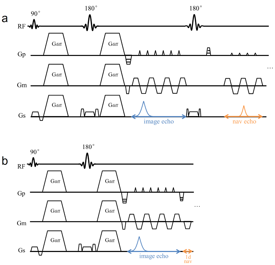
Pulse sequence The proposed multi-shot DWI sequence is shown as in Fig. 1b (compared to the conventional 2-echo acquisition method in Fig. 1a), in which the 1D navigator is acquired right after the image echo at ky=0 for each shot. Compared to the 2-echo acquisition method, the second 180° refocusing pulse and 2nd spin-echo acquisition is saved.
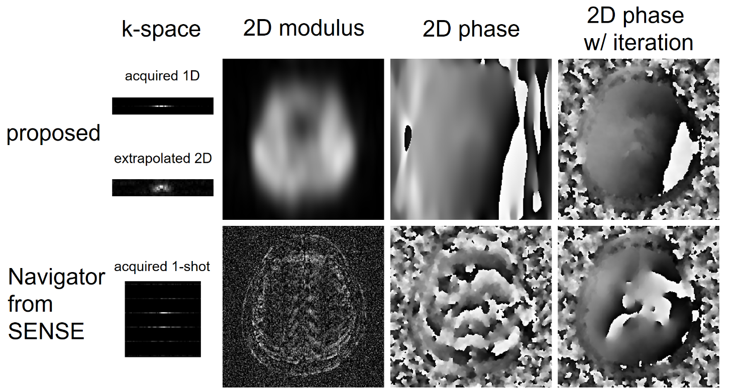
Reconstruction The reconstruction is performed as follows (Fig. 2): 1) The 1D navigator k-space data is first extrapolated to 2D (in this work to 7 ky lines) using GROWL5. The extrapolation coefficients can be calculated from fully-sampled b = 0 data. 2) The coarse initial navigator (net motion-induced phase) for each shot is obtained by subtracting the phase (complex multiplication) of 2D b = 0 images from the 2D diffusion-encoded images (both 2D after k-space extrapolation in step 1). 3) Initial diffusion image is calculated using the coarse initial navigators and image-echo data by the POCSMUSE algorithm4. 4) The diffusion image and motion-induced navigator phases are updated iteratively by POCS using POCSENSE and POCSMUSE as in ref6.
Experiment To demonstrate the advantages of the proposed method, 12-shot diffusion EPI data were acquired on a healthy volunteer on Philips 3T Achieva scanner with an 8-channel head coil. FOV=236×236mm2, voxel size=1×1×4mm3, TE/TR=67/2500ms, 12 diffusion directions with b=1000s/mm2. The acquisition time for the proposed sequence was 6:40 mins for 18 slices. Note that for the conventional 2-echo sequence it would take 8:44 mins with the same parameters. Conventional POCSMUSE (not using the 1D navigator), conventional 1D correction (using the 1D navigator but without extrapolation) and single-shot reference were compared with the proposed method.
Results and Discussion
Fig. 3 shows the results of phase estimation (which is further used in POCSMUSE correction) of a representative shot using the proposed method (1D acquired, 2D extrapolated and with iteration) and POCSENSE method. The SENSE-based navigator shows strong artifacts from R = 12 using the 8-channel coil, while the proposed method shows no significant artifacts. Fig. 4 shows the final diffusion images of a representative slice corrected using POCSMUSE, conventional 1D correction and the proposed method, with or without iterative phase updates. POCSMUSE results show residual aliasing artifacts, which are believed to result from inaccurate phase estimation from high R SENSE. Conventional 1D phase correction cannot handle the phase correctly for nonlinear terms and shows artifacts. The proposed method shows images with reduced artifacts and similar contrast with the single-shot reference, but with higher resolution.
The proposed method kept the high efficiency property of navigator-free multi-shot acquisitions compared to the 2-echo sequences. In this work, the 1D navigator took about 1% more acquisition time of the navigator-free scan. The key idea of the reconstruction is that 2D phase information is obtained by utilizing 1D navigator and parallel imaging technique using multiple coil information, and further updated in the iteration under the assumption that each shot shares the same image intensity (but different motion-induced phases). This method removes the limitation of MUSE-like methods when high R undermines calculated navigators, as we used data set acquired by 12-shot diffusion EPI with 8-channel head coil. This technique can also be further combined with other advanced diffusion acquisition strategy like simultaneous multi-slice imaging.
Conclusion
The proposed multi-shot diffusion imaging using 1D navigator acquisition with parallel imaging and iterative reconstruction can effectively correct the motion-induced nonlinear phase errors in an efficient acquisition way.Acknowledgements
No acknowledgement found.References
1. Porter DA, Heidemann RM. High resolution diffusion-weighted imaging using readout-segmented echo-planar imaging, parallel imaging and a two-dimensional navigator-based reacquisition. Magn. Reson. Med. 2009;62:468–475. doi: 10.1002/mrm.22024.
2. Jeong HK, Gore JC, Anderson AW. High-resolution human diffusion tensor imaging using 2-D navigated multishot SENSE EPI at 7 T. Magn. Reson. Med. 2013;69:793–802. doi: 10.1002/mrm.24320.
3. Ma X, Zhang Z, Dai E, Guo H. Improved multi-shot diffusion imaging using GRAPPA with a compact kernel. Neuroimage 2016;138:88–99. doi: 10.1016/j.neuroimage.2016.05.079.
4. Chen N kuei, Guidon A, Chang HC, Song AW. A robust multi-shot scan strategy for high-resolution diffusion weighted MRI enabled by multiplexed sensitivity-encoding (MUSE). Neuroimage 2013;72:41–47. doi: 10.1016/j.neuroimage.2013.01.038.
5. Lin W, Huang F, Li Y, Reykowski A. GRAPPA operator for wider radial bands (GROWL) with optimally regularized self-calibration. Magn. Reson. Med. 2010;64:757–766. doi: 10.1002/mrm.22462.
6. Zhang Z, Zhang B, Li M, Liang X, Chen X, Liu R, Zhang X, Guo H. Multishot Cartesian turbo spin-echo diffusion imaging using iterative POCSMUSE Reconstruction. J. Magn. Reson. Imaging 2016. doi: 10.1002/jmri.25522.
Figures