3330
Combination of Variable and Constant Flip Angles for Hyperpolarized 129Xe Multi-b Diffusion MRI in a Single Breath-hold1Key Laboratory of Magnetic Resonance in Biological Systems, State Key Laboratory of Magnetic Resonance and Atomic and Molecular Physics, National Center for Magnetic Resonance in Wuhan, Wuhan Institute of Physics and Mathematics, Chinese Academy of Sciences, Wuhan, People's Republic of China, 2University of Chinese Academy of Sciences, Beijing, People's Republic of China
Synopsis
To propose a novel flip angle scheme for hyperpolarized gas multi-b diffusion MRI. It combined the variable and constant flip angles and was named as the combination of variable and constant flip angle (CVCFA). Computer simulation was used to systematically compare the proposed flip angle scheme with the common-used flip angle schemes, including the interleaved constant flip angle (ICFA) and the variable flip angle (VFA) schemes. The CVCFA scheme was used for hyperpolarized xenon diffusion MRI to measure the pulmonary morphology in rats, noninvasively. The results showed the CVCFA was suited for the hyperpolarized gas multi-b diffusion MRI.
PURPOSE
To propose a novel flip angle scheme for multi-b diffusion MRI with hyperpolarized (HP) gas in a single breath-hold, which was used to measure the pulmonary morphology in vivo and noninvasively.METHODS
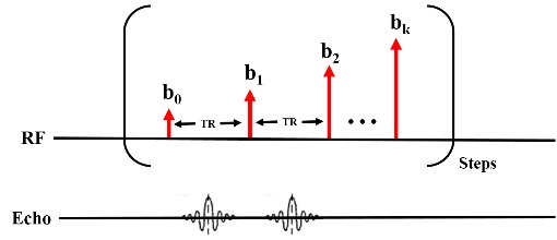
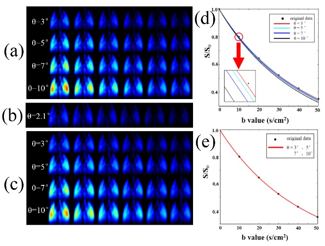
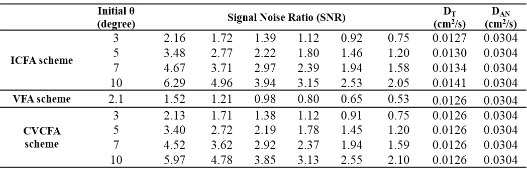
Computer simulation was firstly used to systematically evaluate the influence of the common-used flip angle scheme and the proposed scheme in HP gas multi-b diffusion MRI. Interleaved constant flip angle (ICFA) and the variable flip angle (VFA) schemes are the currently used methods. Our proposed method combined the variable and constant flip angle schemes and was named as the combination of variable and constant flip angle (CVCFA). The diagram of CVCFA was shown in the Fig.1, where k represented the numbers of b value and ‘Steps’ represented the phase encoding. According to the theory proposed by Yablonskiy et al, the diffusion of HP gas in lungs is anisotropic and can be described with the equation [1]: S=S0exp(-bDT)[π/(4bDAN)]1/2∅[(bDAN)1/2], where S are images with diffusion gradient, S0 is the image without diffusion gradient, DT represents the transverse diffusion coefficient, DAN represents the anisotropy of diffusion. ∅ is the error function. In the simulation, we set the DT= 0.0126 cm2/s and DAN=0.0304 cm2/s, according to our previous paper. The number of b was six (0, 10, 20, 30, 40, 50 s/cm2) and the phase encoding steps was 128. The results from the three schemes were compared and analyzed, including the image SNR and accuracy. Afterwards, the CVCFA scheme was used for HP xenon multi-b diffusion MRI in a single breathe-hold, to measure the pulmonary morphology in four healthy Wistar male rats (230±20 g). Four b values (0,10,20,30,40 s/cm2) were used and the diffusion time was 2 ms in the experiment.RESULTS and DISCUSSION
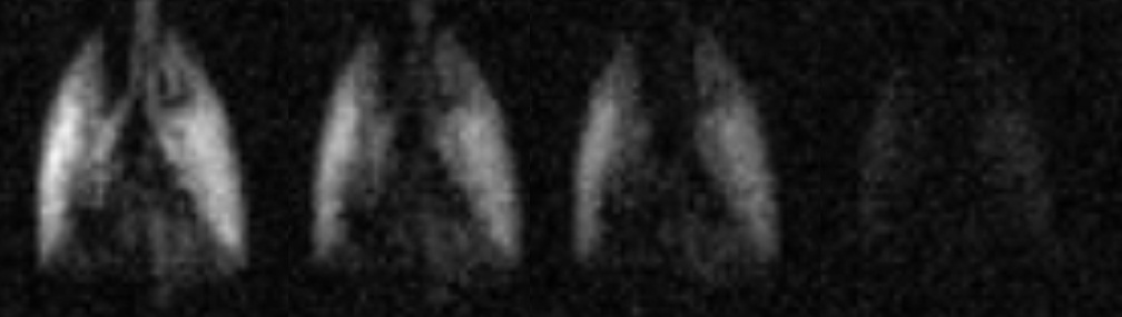
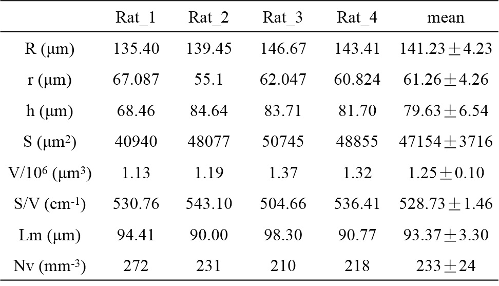
Computer simulation: Fig.2 and Table.1 showed the simulation results with different flip angle schemes. Fig.2 (a) was the result of ICFA scheme with different flip angles (3°, 5°, 7°and 10°). Although the image SNR increased with the increasing flip angle, the fitting curve deviated from the original data more largely, as illustrated in the Fig.2 (d). The deviation would induce the error of fitting results. Table.1 showed that the DT from the ICFA scheme were different from the preset value 0.0126 cm2/s and the error increased with the increasing flip angle. Fig. 2 (b) showed the result of VFA scheme. According to the previous theory, the initial flip angle depends on the number of RF pulses in VFA scheme. Although the fitting curve could overlap the original data as shown in Fig.2 (e), the image SNR was very low. Fig.3 (c) showed the result of CVCFA scheme with different flip angles (3°, 5°, 7°and 10°). With the same initial flip angle, the images SNR obtained by ICFA scheme were nearly the same with those obtained by CVCFA scheme. Meanwhile, the Fig.2 (e) shows that the fitting curve from CVCFA scheme could overlap the original data completely. Table.1 also showed that the DT and DAN were consistent with the preset values. In vivo experiment: Fig.3 shows the representative multi-b lung images obtained in a single breath-hold with the CVCFA scheme. From left to right, the b values were 0, 10, 20, 30 s/cm2, respectively. Although no filtering process was applied, the image quality was still good. The image SNR were 24.00, 18.91, 13.77 and 11.15 from left to right, respectively. The morphologic parameters of lung microstructure were summarized in Table.2, including the outer acinar airway radius (R), the internal acinar airway radius (r), the depth of acinar (h) et al. The results were consistent with those reported in the previous studies [2,4,5]. In this study, the application of different flip angle schemes for the multi-b HP gas diffusion MRI were first systematically analyzed and compared. The traditional VFA scheme was accurate for the multi-b data fitting, but image SNR was low. The ICFA scheme could be used to obtain the high SNR images, but the accuracy of the fitting results will be effected, especially the DT value. The proposed CVCFA scheme combined the advantage of the two traditional flip angle schemes, and could obtain accurate images as well as high SNR for multi-b HP gas diffusion MRI in one breath-hold.Acknowledgements
We acknowledge the support by the National Natural Science Foundation of China (81227902, 81625011) and National Program for Support of Eminent Professionals (National Program for Support of Top-notch Young Professional).References
1. Sukstanskii AL,Yablonskiy DA. Lung morphometry with hyperpolarized 129Xe: theoretical background. Magn Reson Med 2012; 67: 856-866.
2. Ruan W, Zhong J, Wang K, Wu G, Han Y, Sun X, Ye C, Zhou X. Detection of the mild emphysema by quantification of lung respiratory airways with hyperpolarized xenon diffusion MRI. J Magn Reson Imaging 2016; doi: 10.1002/jmri.25408.
3. Deng H, Zhong J, Ruan W, Chen X, Sun X, Ye C, Liu M, Zhou X. Constant-Variable Flip Angles for Hyperpolarized Media MRI. J Magn Reson 2016;263: 92-100.
4. Ouriadov A, Fox M, Hegarty E, Parraga G, Wong E, and Santyr GE. Early stage radiation-induced lung injury detected using hyperpolarized Xe Morphometry: Proof-of-concept demonstration in a rat model. Magn Reson Med 2016; 75 :2421-2431.
Figures