3324
Single Breath CSSR-DWI: A New Method to Simultaneously and Quantitatively Assess the Changes of Respiratory Membrane and Pulmonary Microstructure in Human1Key Laboratory of Magnetic Resonance in Biological Systems, State Key Laboratory of Magnetic Resonance and Atomic and Molecular Physics, National Center for Magnetic Resonance in Wuhan, Wuhan Institute of Physics and Mathematics, Chinese Academy of Sciences, Wuhan, People's Republic of China, 2Department of Radiology, Zhongnan Hospital of Wuhan University, Wuhan, People's Republic of China
Synopsis
The blood-gas exchange function and the pulmonary microstructure are generally affected by the lung inflation levels. Here We developed a new method Single Breath CSSR-DWI to simultaneously quantify the respiratory membrane and the pulmonary microstructure via hyperpolarized 129Xe in a single breath. A new parameter SVRd/g was defined from ADC and SVRd to characterize the ‘%-predicted dissolved SVR’. Human pulmonary functional and structure information were successfully obtained in a single breath via hyperpolarized 129Xe. Compared to the healthy young subjects, SVRd/g of the asymptomatic aged subjects decreases while the size of the pulmonary microstructure increases.
Introduction
The functional information of the pulmonary blood-gas exchange could be obtained by the chemical shift saturation recovery (CSSR) method and related xenon uptake model1. Meanwhile, hyperpolarized xenon diffusion weighted MR imaging (DWI) could offer the information of pulmonary microstructure2. However, the blood-gas exchange function and the pulmonary microstructure are generally affected by the lung inflation levels3. Therefore, simultaneously acquiring CSSR data and DWI data in a single breath is necessary to obtain the accurate pulmonary physiological parameters. In this study, we developed a new method, named as Single Breath CSSR-DWI, to quantify the respiratory membrane and the pulmonary microstructure via hyperpolarized 129Xe in a single breath.Methods
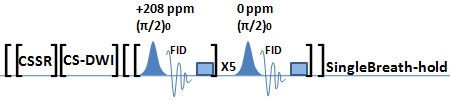
The pulse sequence used in this study was shown in Fig. 1 (a total time of 10 s). For the CSSR, three 1.6 ms Gaussian saturation RF pulses with the flip angle of 90° (90° phased, centered at +208 ppm relative to the resonance frequency of xenon in alveoli) separated by gradient spoilers were used to destroy the dissolved xenon. After a series of variable delay that controlled how much xenon gas can enter the lung parenchyma and the blood from the alveolar airspaces, a 1.2 ms Gaussian excitation RF pulse with the flip angle of 90° (0° phased, centered at +208 ppm) was applied for data collecting (TE=0.7 ms, Bandwidth=10 kHz, 512 data points). The process was repeated 21 times in the same breath hold with delay times ranging from 5 ms to 700 ms (a total time of 5.3 s). For the weighted images of DWI, b = 12 s/cm2 (bipolar diffusion gradients, the diffusion time Δ is 3.6 ms, the ramp time τ is 0.3 ms, no gaps between the bipolar gradients, 5 slices) and the compressed sensing (CS) method4 was used to accelerate the acquisition (a total time of 4.2 s). After acquiring the DWI data, spectra were collected to calibrate the influence of the RF pulse centered at dissolved xenon resonance frequency on gas xenon (a total time of 0.5 s). All the experiments were performed on a 1.5 T whole-body MRI Scanner (Avanto, Siemens Medical Solutions) using a homebuilt transmit-receive vest RF coil. Enriched xenon (86% 129Xe) was polarized using a homebuilt xenon polarizer. One liter of xenon mixture (30% xenon + 70% N2) was inhaled from functional residual capacity before each measurement. Informed consent was obtained from each of 4 healthy young subjects (aged between 23 and 27, HY) and 3 asymptomatic aged non-smoking subjects (aged 63-85 years, HA). The protocol was approved by the institutional review board (IRB). Five parameters were obtained by fitting CSSR data to MOXE: SVRd, d, ξ/d, Hct, tX. A new parameter SVRd/g was defined from ADC and SVRd to characterize the ‘%-predicted dissolved SVR’. SVRd/g = SVRd/(4/(2(Δ+δ)ADC)0.5), where δ is the duration time of the diffusion gradients and ADC the mean apparent diffusion coefficient of the whole lung.Results
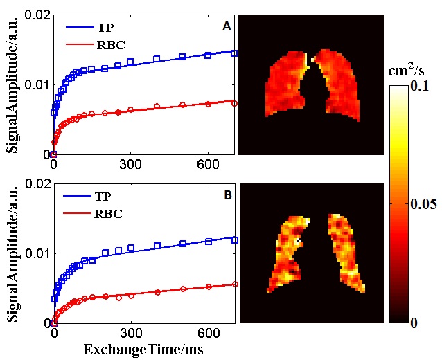
For the HY subjects, SVRd/g = 1.24 ± 0.05, ADC = 0.040 ± 0.002 cm2/s. For the HA subjects, SVRd/g = 0.77 ± 0.01, ADC = 0.052 ± 0.005 cm2/s. Figure 2 showed the CSSR curves and the ADC maps of the third slice of HY4 and HA1. Compared to HY subjects, SVRd/g of HA subjects significantly decreased (p<0.05) while ADC significantly increased (p<0.05).Conclusion
Human pulmonary functional and structure information were successfully obtained in a single breath via hyperpolarized 129Xe. The parameters obtained from the MOXE model agreed well with the previous study5, and the changes of ADC were in agreement with the results reported by Kaushik6. Compared to the healthy young subjects, the ‘%-predicted dissolved SVR’ of the asymptomatic aged subjects decreases while the size of the pulmonary microstructure increases.Acknowledgements
We acknowledge the support by the National Natural Science Foundation of China (81227902, 81625011, 81601491) and National Program for Support of Eminent Professionals (National Program for Support of Top-notch Young Professionals).References
1. Li H et al. Magn Reson Med 2016; 76: 408-416.
2. Ruan W et al. J Magn Reson Imaging 2016.
3. Dregely I et al. Magn Reson Med 2012; 67: 943-953.
4. Lustig M et al. Magn Reson Med 2007; 58: 1182-1195.
5. Chang Y et al. Magn Reson Med 2014; 71: 339-344.
6. Kaushik et al. Magn Reson Med 2011; 65: 1155-1165.
Figures