3316
Analysis of Regional Lung Function Detected by Hyperpolarized Xenon-129 MRI in Subjects with Interstitial Lung DiseasesKun Qing1, Talissa A. Altes2, John P. Mugler, III1, Nicholas J. Tustison1, Kai Ruppert3, Jaime F. Mata1, Yun Michael Shim1, G.Wilson Miller1, Iulian C. Ruset4, F.William Hersman4,5, and Borna Mehrad1
1University of Virginia, Charlottesville, VA, United States, 2University of Missouri School of Medicine, Columbia, MO, United States, 3University of Pennsylvania, Philadelphia, PA, United States, 4Xemed, LLC, Durham, NH, United States, 5University of New Hampshire, Durham, NH, United States
Synopsis
Previous study showed that hyperpolarized xenon-129 MRI is highly sensitive in detecting functional changes in lungs with interstitial lung diseases (ILD). The degree to which these changes vary regionally in the lung has not been determined, however. In this work, we compared abnormalities in lung function in different regions of the lung, and found significant differences in xenon-129 gas uptake between subjects with ILD and controls. These results support that xenon-129 MRI may provide unique information about lung physiology associated with lung fibrosis.
Introduction
In our preliminary study using hyperpolarized xenon-129 (Xe129) MRI [1], we found subjects with the histologic or radiographic diagnosis of usual interstitial pneumonia (UIP) had higher gas uptake by lung tissue (tissue-to-gas ratio) and lower gas transfer from tissue to red blood cells (RBCs, RBC-to-tissue ratio) as compared to controls. However, the extent to which the abnormalities varied in different areas of the lung has not been determined.Purpose
The purpose of this work is to evaluate regional alterations of lung function detected by Xe129 MRI and compare it with regional information of the lung acquired by CT.Methods
Ten subjects with UIP (age 66±15) and ten healthy controls (age 58±14) were recruited for this study and underwent Xe129 MRI and CT. Two functional measures were produced from the Xe129 MRI results, including tissue-to-gas and RBC-to-tissue ratios [2]. CT Hounsfield (HU) data, reflecting lung tissue density, was used for comparison. Two segmentation methods were used to analyze the regional differences of measures by MRI and CT. First, lobar segmentation [3], by which lungs were segmented into five lobes, was used to compare the differences between upper and lower lobes of the lung. And segmentation of outer rind/inner core (within/beyond 10 mm of the lung boundary) [4] was used to compare differences of function in central and distal parts of the lung. Differences of Xe129 tissue-to-gas, RBC-to-tissue ratios, and for CT HU data between upper lobes and lower lobes (Dul = mean values in upper lobes - mean values in lower lobes), and between outer rind and inner core (Doi = mean values in the outer rind - mean values in the inner core) were calculated for each individual subject. And the results of UIP subjects and controls were compared using student’s t-test.Results
Representative segmentation masks were shown in Fig. 1 (left: lobar segmentation, right: segmentation of outer rind/inner core). In this work, we did not find any differences in Dul and Doi of CT HU between the two groups (CT HU, Dul: healthy: -58+33, IPF: -71+52, P= 0.53, Fig. 2a; Doi: healthy: 80+55, IPF: 104+36, P=0.27, Fig. 2b). However, the UIP subjects showed significantly higher Dul of gas uptake by tissue (tissue-to-gas ratio, Dul: healthy: -0.15%+0.15%, IPF: 0.13%+0.13%, P<0.001, Fig. 2c), and higher Dul of gas transfer from tissue to RBCs (RBC-to-tissue ratio, Dul: healthy: -0.017+0.044, IPF: 0.030+0.040, P=0.026, Fig. 2e) than controls. UIP subjects also showed a trend towards higher Doi in gas transfer from tissue to RBCs in the distal lungs (RBC-to-tissue ratio, Doi: healthy: -0.057+0.041, IPF: -0.014+0.055, P=0.06, Fig. 2f) than healthy subjects. In contrast, the the Doi of tissue-to-gas ratios did not differ significantly between the groups (Doi: healthy: 0.01%+0.05%, UIP: -0.00%+0.08%, P=0.53, Fig. 2d).Discussions and Conclusion
In this study, by using two imaging segmentation tools, we performed analysis of regional lung function in subjects with UIP and compared the results with those from controls. An interesting finding is that in controls, upper lobes of the lungs had lower tissue gas uptake and gas transfer from tissue to RBCs than lower lobes (mean Dul= -0.15% for tissue-to-gas ratios, and -0.017 for RBC-to-tissue ratios). But in subjects with UIP, this was reversed (mean Dul= 0.13% for tissue-to-gas ratios and 0.03 for RBC-to-tissue ratios). While no regional differences in CT HU were found in this study. These could be due to the higher sensitivity of Xe129 MRI to detect alterations of lung function. The extent to which these functional data are related to changes of lung physiology in interstitial lung diseases awaits further investigation.Acknowledgements
This work was supported by a Transformative, Collaborative Science Pilot Grant from University of Virginia School of Medicine and NIH R01 HL109618.References
[1] Qing K et al. Proc of ISMRM 24 (2016): 1147. [2] Qing, K., et al. J Magn Reson Imaging, 2014. 39(2): p. 346-59. [3] Tustison NJ et al. Magn Reson Med, 2016. 76(1): 315-20. [4] Tustison NJ et al. Magn Reson Med 2010. 63(6): 1448-1455.Figures
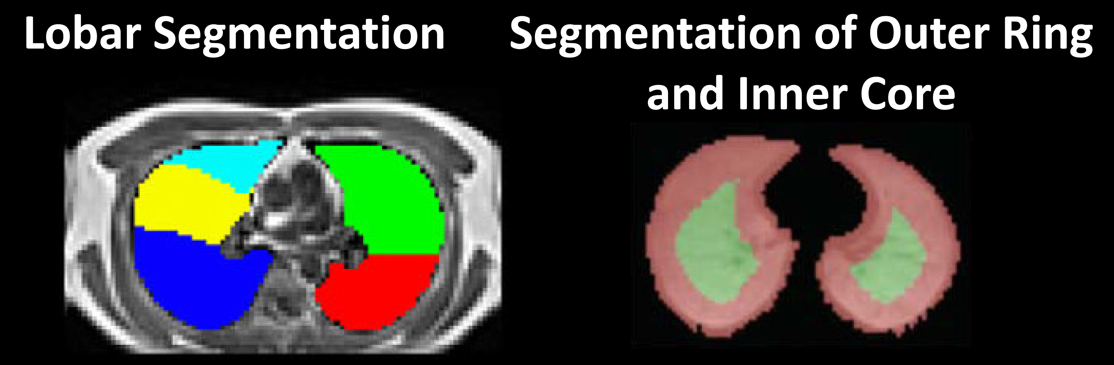
Representative lung segmentation masks for two segmentation methods in
transversal views: left: lobar segmentation, right: segmentation of outer rind
and inner core. Outer rind and inner core were defined as within and beyond
10mm of lung boundary, respectively.
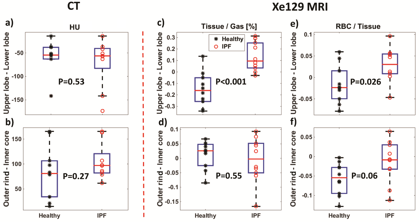
Differences of Xe129
tissue-to-gas, RBC-to-tissue ratios, and CT HU data between upper lobes and
lower lobes (mean values in upper lobes - mean values in lower lobes), and
between outer rind and inner core (mean values in the outer rind - mean values
in the inner core) were compared between the UIP and healthy groups.