3312
MR Elastography vs. Diffusion-weighted MRI in Characterization of Anterior Mediastinal Solid Tumours1Diagnostic Radiology, Cancer Hospital, Chinese Academy of Medical Sciences, Peking Union Medical College, Beijing, People's Republic of China
Synopsis
Anterior mediastinal solid tumours were routinely interpreted on CT and MR imaging. However, imaging features of these tumours could be non-specific; evidence suggestive of definitive diagnosis was needed and obtained with invasive procedure of biopsy for some cases. In the present study, we attempted to extent the application of MR elastography to the mediastinum in characterization of anterior mediastinal solid tumours, with the comparison to diffusion-weighted MRI. Thirty-four patients histologically-confirmed with thymic carcinoma in 10, thymoma in 10 and lymphoma in 14 were evaluated. It was found that stiffness value measured on elastogram was significantly higher in thymic carcinoma than that of thymoma and lymphoma. However, measurements of apparent diffusion coefficient (ADC) were not significantly different among three groups. It was demonstrated that MR elastography reflecting the mechanical properties of tumours can be used to characterize anterior mediastinal solid tumours, particularly in distinguishing thymic carcinoma from lymphoma.
Purpose
In this preliminary study, we proposed an investigation on the
feasibility of MR elastography in characterization of the mechanical properties
of anterior mediastinal solid tumours in relation to the histopathology, and with
the comparison to diffusion-weighted MRI.
Methods
Institutional review board approval and informed consent were
obtained. Thirty-four patients (18 men, 16 women; age range 23-69 years, mean
46.5 years) with anterior mediastinal solid tumours were enrolled. All the
patients were examined with MR examination consisting of diffusion-weighted
imaging (DWI) and MR elastography on a 3.0 T whole body scanner. A
manually-drew regions of interest (ROI) covering as large as the areas of the
tumour was placed on DWI image on which the largest axial diameter of tumours
was shown. The identical ROI then was copied to the corresponding elastogram at
the same level for each lesion. The measurements of apparent diffusion
coefficient (ADC) value on DWI and stiffness value on elastogram were
automatically obtained. All measurements were performed three times and the
average values were recorded. Two-way ANOVA test was used to examine the
difference of ADC and stiffness values with the correlation of pathological
results. Receiver operating characteristic (ROC) curve analyses were performed
to determine the area under curve (AUC) for accuracy of MR elastography and DWI
in differential diagnosis.
Results
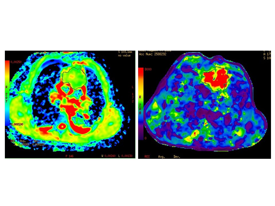
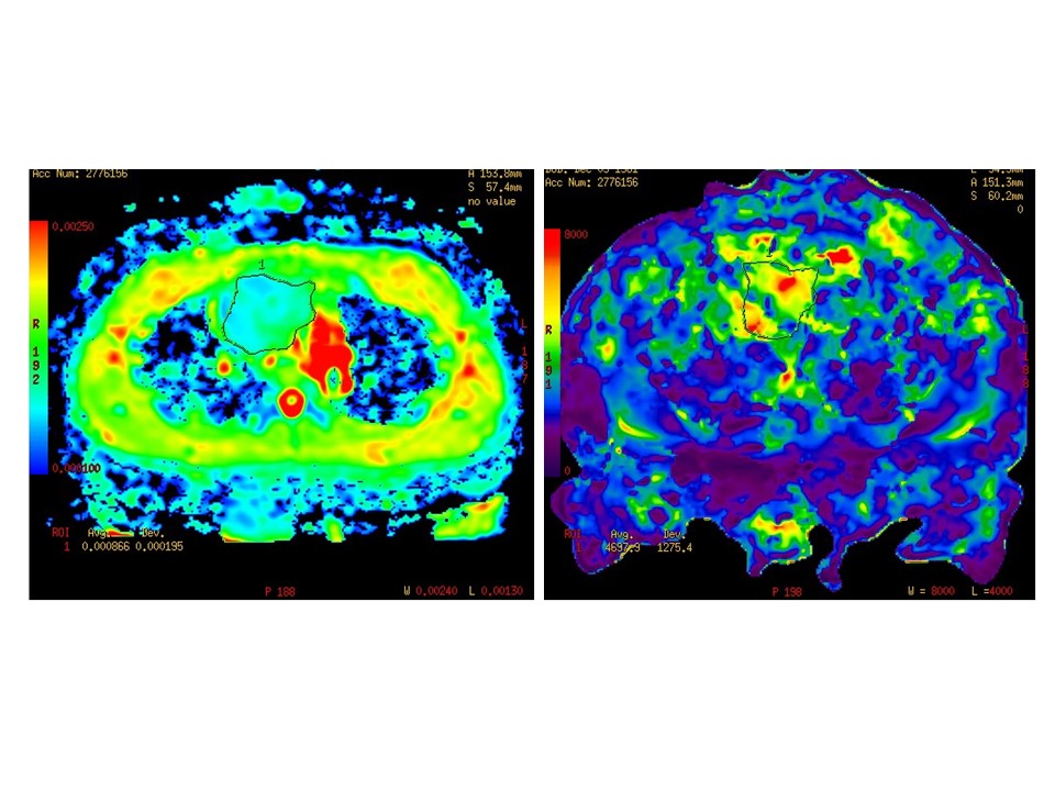
Those 34 patients with anterior mediastinal tumours were histologically confirmed with thymic carcinoma in 10, thymoma in 10, and lymphoma in 14. Higher stiffness value of thymic carcinoma was measured as 6.56±1.03 kPa, followed by 4.81±0.95 kPa for thymoma, and 3.89±1.23 kPa for lymphoma. Stiffness value of thymic carcinoma was significantly higher than that of thymoma (p = 0.006) and lymphoma (p = 0.000). However, ADC values were not significantly different among those three groups, with the measurement of ADC being 1.33±0.25 (×10−3mm2/s) in thymic carcinoma, 1.28±0.53 (×10−3mm2/s) in thymoma, and 1.26±0.48 (×10−3mm2/s) in lymphoma, respectively.
Results of ROC analysis for stiffness values showed that AUC for distinguishing thymic carcinoma from thymoma and lymphoma was 0.917. The feasible threshold value was 4.84 kPa with a sensitivity of 100% and specificity of 66.67% in diagnosis of thymic carcinoma (Figure 1). AUC for stiffness values in distinguishing lymphoma from thymic carcinoma and thymoma was 0.843. The feasible threshold value was 4.08 kPa with a sensitivity of 71.43% and specificity of 90.7% in diagnosis of lymphoma (Figure 2).
Discussion
To the best of our knowledge, the present study is the first in the literature that verify the capability of MR elastography in characterization the anterior mediastinal solid tumours using elasticity mapping.
Those patients diagnosed with thymic carcinoma in our study were all squamous cell carcinoma, which is histologically characterized by abundant large epithelial cells arranged in nests or cords, and the broad zone of fibrohyaline stroma separating the tumour cell nests. (1) It was hard and firm and supposed to be with higher stiffness evaluated on MR elastography. On the other hand, lymphoma was usually demonstrated as diffused pattern of tumour cells separated with irregular collagen fibris and fibers, (1) which could be assessed on MR elastography with lower stiffness. The results of our study showed that MR elastography can be considered as an effective technique to differentiate thymic carcinoma from lymphoma.
Previous reports have proved that DWI could be used to distinguish malignancies from benign tumors as significantly lower ADC values were revealed for malignant tumours. (2, 3) In the present study, however, measurements of ADC values were not significantly different among three groups of tumours. The superior performance of MR elastography in differential diagnosis of anterior mediastinal solid tumours is likely due to the fact that MR elastography measures the mechanical property of tissue, whereas ADC values measured on DWI represents diffusion that could be affected by several factors, including T2, perfusion, cellularity and possibly prandial state. (4)
Conclusion
The study demonstrates the feasibility of MR elastography in the characterization of stiffness of anterior mediastinal solid tumours, with the superiority in distinguishing thymic carcinoma from lymphoma when compared with DWI.Acknowledgements
No acknowledgement found.References
1. WHO Classification of Tumours of the Lung, Pleura, Thymus and Heart. Lyon, France: World Health Organization; 2004.
2. Seki S, Koyama H, Ohno Y, et al. Diffusion-weighted MR imaging vs. multi-detector row CT: Direct comparison of capability for assessment of management needs for anterior mediastinal solitary tumours. Eur J Radiol. 2014 May; 83(5): 835-42.
3. Gümüstas S, Inan N, Sarisoy HT, et al. Malignant versus benign mediastinal lesions: quantitative assessment with diffusion weighted MR imaging. Eur Radiol. 2011 Nov; 21(11):2255-60.
4. GH Jajamovich,H Dyvorne,C Donnerhack, B Taouli. Quantitative liver MRI combining phase contrast imaging, elastography, and DWI: assessment of reproducibility and postprandial effect at 3.0 T. Plos One. 2014, 9 (5):e97355.
Figures