3306
Arterial Spin Labeling Imaging for Evaluation of Renal Changes in Remaining and Donated Kidneys Early after Living Renal Allograft Transplantation1Department of Radiology, Tianjin First Center Hospital, TianJin, People's Republic of China, 2Organ Tranplant Center, Tianjin First Center Hospital, TianJin, People's Republic of China
Synopsis
Arterial Spin Labeling (ASL) MRI is a noninvasive approach for assess renal function,which provides a quantitative measure of perfusion without the use of an exogenous contrast. Our study detected cortex perfusion changes in remaining and donated Kidneys. The result showed ASL can be used for detecting renal changes in remaining and donated kidneys early after living renal allograft transplantation.
Target audience
Researchers and clinicians with an interest in functional magnetic resonance imaging of kidney.Introduction
Kidney transplantation has become the most successful treatment option for patients with end-stage renal diseases,which,in turn,increases the demand for living kidney donation.However,donors experience reduced renal function along with an increased risk of proteinuria and hypertension;for transplant recipients,the major long-term risk is allograft failure,with the majority failing before the end of a recipients’ lifetime.Arterial Spin Labeling (ASL) MRI is a noninvasive approach for assess renal function,which provides a quantitative measure of perfusion without the use of an exogenous contrast.Renal ASL MR imaging has shown good reproducibility in healthy subject,transplant recipients,and patients with chronic kidney disease(CKD).Our study aims to evaluate ASL MR imaging data from the same living kidney donors and their recipients to determine potential renal perfusion changes associated with uninephrectomy and transplantation in remaining and transplanted kidneys.Methods
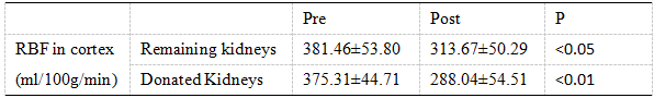
Ten kidney donors(mean age 52.0±4.6 years, 5 females and 5 males) and ten matched allograft recipients (mean age 37.2±5.1 years, 1 females and 9 males)were recruited.Donors with history of renal diseases, hypertension and other vascular diseases,and abnormal findings in kidney on MRI were excluded from the study.All allograft recipients received routine triple immunosuppression therapy after transplantation. All donors underwent a MR examinations prior to and early after uninephrectomy,and recipients underwent MR examinations only early after transplantation.All MR examinations in this study were performed on a clinical 3.0T MR scanner(Magnetom Trio;Siemens,Erlangen,Germany).For quantitative assessment of the renal perfusion,the flow sensitive alternating inversion recovery-true fast image with steady state precession(FAIR-TrueFISP) sequence was performed.The oblique-sagittal ASL was obtained with following parameters: TE/TR 2.24/4.48ms, flip angle 70°,voxel size 2.3×2.3×5.0mm3,slice thickness 5 mm,number of slices 1,inversion time(TI) 1200ms,measurements 12.For donors,respiratory-triggered acquisition was used to weaken the impact of respiratory motion. The renal blood flow (RBF) values were measured only in the cortex in this study1.Three sections nearest to the renal hilum were selected for region of interest (ROI) analysis.For each selected section,one ROI of 80-120 pixels was manually delineated to cover the renal cortex.The parameters of cortex were compared between before and after operations by using Paired Student t-test, performed with SPSS 19.0 software (SPSS Inc. Chicago, IL, USA) and results with P values less than 0.05 were considered statistical significant.
Results and Discussion
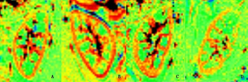
Table 1 summerizes mean values of RBF parameters in donors and recipients. Mean RBF values in the cortex of remaining kidney post-nephrectomy was lower than pre-nephrectomy. Mean RBF in donated kidneys reduced early after transplantation.This result was similar to the previous study2,3. For donors,one possible explanation of the finding is that hypertrophy of he naive kidney result in lower perfusion because the unit of measurement is normalized to mass,and implicity volume,of tissue. The decline of cortical perfusion in donated kidneys may result from calcineurin-inhibitor-induced tubular dysfunction.Figure 1 illustrates image examples of RBF of kidneys in a donor and a recipient.Conclusion
ASL was able to reflect renal perfusion changes associated with uninephrectomy and transplantation in remaining and transplanted kidneys.ASL can be used for detecting renal changes in kidney donors and allograft recipients ,and monitoring renal function noninvasively.Acknowledgements
References
[1]Artz NS,Sadowski EA,Wentland AL,et al.Reproducibility of renal perfusion MR imaging in native and transplanted kidneys using non-contrast arterial spin labeling.J Magn Reson Imaging 2011;33(6);1414-21.[2] Marica C, Rachel H,Jonathon O, et al. Renal blood flow using arterial spin labelling MRI and calculated filtration fraction in healthy adult kidney donors Pre-nephrectomy and post-nephrectomy. European Radiology 2015;25(8):2390–6. [3]Eisenberger U, Binser T, Thoeny HC,et al.Living renal allograft transplantation:diffusion-weighted MR imaging in longitudinal follow-up of the donated and the remaining kidney.Radiology 2014;270 (3):800-8.Figures