3297
Prospective study of intravoxel incoherent motion (IVIM) MRI of bladder as a biomarker for prediction of bladder cancer aggressiveness1Department Of Imaging Diagnosis, National Cancer Center/Cancer Hospital, Chinese Academy of Medical Science and Peking Union Medical College, Beijing, People's Republic of China, 2China Rehabilition Research Center, 3GE Healthcare, MR Research China, Beijing China
Synopsis
Comparing the IVIM-MRI parameters with postoperative histopathological findings, we found significant correlations between quantitative parameters derived from IVIM-MRI and histological grade, as well as the depth of invasion. We concluded that IVIM-MRI quantitative parameter could be a promising imaging biomarker for prediction of bladder cancer aggressiveness.
Purpose
As we all know, apparent diffusion coefficient (ADC) value derived from traditional diffusion-weighted MRI (DW-MRI) has been considered useful for pathological staging and histological grading in bladder cancer. To our knowledge, no study has been done to explore whether IVIM MR imaging is useful in the evaluation of pathological staging and histological grading in bladder cancer. Our study was to evaluate the association of IVIM-MRI quantitative parameters with the histological grade and the depth of invasion in bladder cancer.
Materials and Methods
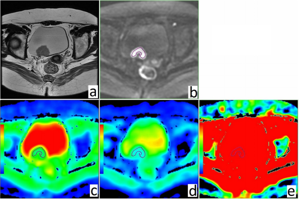
Twenty-three patients (19 men, 4 women, mean age 60 years) were enrolled in this study and inspected through conventional MR and IVIM-MR examination with a 3.0-T MR scanner (GE Discovery MR750 with an 8-channel CTL Target Array Coil) from April 2016 to September 2016. In all patients, urothelium carcinoma of bladder was confirmed by surgery and pathology. Research sequences including diffusion-weighted imaging with 12 b values ( 10, 25, 50, 75, 100, 150, 200, 400, 800, 1000, 1500, 2000s/mm2 ) were performed to evaluate diffusion and perfusion characteristics of bladder cancer. IVIM-MRI parameters (standard ADC ADCstandard, slow ADC D, fast ADC D*, and fraction of fast ADC f) of bladder carcinomas were measured by using the FuncTool on GE AW4.6 workstation. Measurements were conducted by two radiologists who were blinded to histopathological results through a region-of-interest from the most hypointensive region of the mass lesions, which were based on the b=1000 s/mm2 image. Comparisons of IVIM-MRI parameters with histopathologic features, which included histologic grade and the depth of muscle invasion, were performed using unpaired t tests. Diagnostic performance was calculated by means of receiver operating characteristics (ROC) statistics.Results
The D and ADCstandard values in high-grade bladder cancer were significantly lower than those in the low-grade (P < 0.05), whereas no significant difference was found in f and D* values. The ROC curve showed that AUC value for D and ADCstandard to distinguish high-grade bladder cancer from low-grade was 0.863 and 0.814, respectively. At the cut-off D value lower than 0.86×10⁻³mm2/s, the high-grade bladder cancer could be differentiated from the low-grade with a sensitivity of 81.3% and specificity of 83.3%, as well as a cut-off ADCstandard value lower than 1.74×10⁻³mm2/s with a sensitivity of 87.5% and specificity of 66.7%. The ADCstandard , f and D values in muscle invasive bladder cancer (MIBC) were significantly lower than those in nonmuscle invasive bladder cancer (NMIBC) (P < 0.05), whereas no significant difference was found in D* value. The ROC curve indicated that AUC values for ADCstandard , f and D value for distinguishing MIBC from NMIBC was 0.873, 0.849 and 0.817, respectively. At the cut-off ADCstandard value lower than 1.15×10⁻³mm2/s, MIBC could be differentiated from the NMIBC with a sensitivity of 75.0% and specificity of 100%, as well as a cut-off f value lower than 0.38 with a sensitivity of 75.0% and specificity of 100%, and a cut-off D value lower than 0.80×10⁻³mm2/s with a sensitivity of 87.5% and specificity of 78.6%.Conclusion
D and ADCstandard values obtained by IVIM-MRI are promising imaging biomarkers for the prediction of bladder cancer histological grade. ADCstandard , f and D values derived from IVIM-MRI have the potential to predict the T staging of bladder cancer. Further studies are necessary to improve these values in guiding of decision making in clinical practice.Acknowledgements
No acknowledgement found.References
[1]Zhou,G. Contrast-enhanced dynamic and diffusion-weighted MR at 3.0T to assess aggressiveness of bladder cancer. Eur J Radiol, 2014, 83(11), 2013-2018.
[2]Avcu,S. The value of diffusion-weighted MRI in the diagnosis of malignant and benign urinary bladder lesions. Br J Radiol, 2011, 84(1006), 875-882.
Figures