3283
Impact of temporal resolution on quantitative renal perfusion MRI: Assessment using a single contrast injection and a continuous golden-angle radial sampling technique with iterative reconstruction.1Department of Clinical Radiology and Nuclear Medicine, University Medical Center Mannheim, Medical Faculty Mannheim, Mannheim, Germany, 2Department of Clinical Radiology and Nuclear Medicine, University Medical Center Mannheim, Medical Faculty Mannheim, Heidelberg University, 3Siemens Healthcare GmbH, Erlangen, Germany, 4Center for Advanced Imaging Innovation and Research (CAI2R), Department of Radiology, New York University School of Medicine, New York, NY, United States, 5Computer Assisted Clinical Medicine, Medical Faculty Mannheim, Heidelberg University, Mannheim, Germany
Synopsis
To evaluate the impact of temporal resolution on quantitative renal perfusion MRI, an intra-individual comparison of retrospectively reconstructed datasets with 4 different temporal resolutions (1.5s to 10.1s) was performed in 22 patients. This was achieved using a continuously acquired sequence that uses a combination of radial sampling, sparse imaging, iterative reconstruction, parallel imaging, and a single contrast injection. No statistically significant differences in renal plasma flow were found between the groups. This suggests that the effect of temporal resolution plays a subordinated role in quantitative renal perfusion MRI.
Purpose
Dynamic contrast-enhanced (DCE) MRI is an established method for the quantitative assessment of renal perfusion1. However, temporal resolution of DCE sequences varies in literature; there is no definite consensus on the minimal temporal resolution required for reliable quantitative perfusion assessment2. Recently, a novel sequence that uses a combination of radial sampling, sparse imaging, iterative reconstruction, and parallel imaging (Golden-angle RAdial Sparse Parallel imaging, GRASP) has been shown to provide both morphological as well as quantitative renal perfusion information from a single acquisition3. However, the impact of different temporal resolutions on quantitative parameters in this setting is still unclear. Thus, the purpose of this study was the in-vivo evaluation of different temporal resolutions for quantitative renal DCE using a single contrast injection.Methods
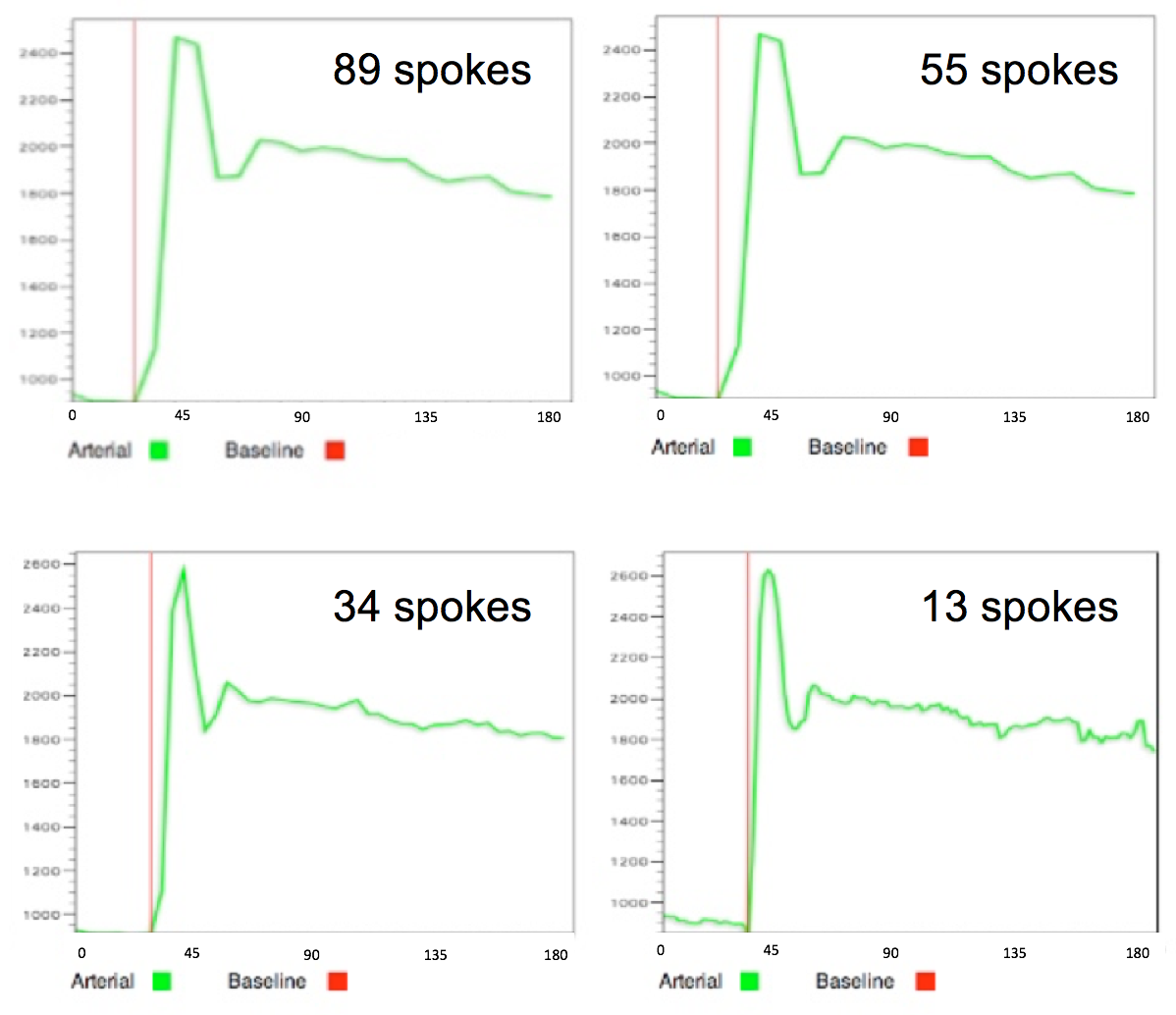
22 patients (7 female; 58.7±21.2 years) underwent imaging with a prototypic fat-suppressed, golden-angle radial stack-of-stars T1-weighted 3D spoiled gradient-echo sequence on a 3 T scanner (MAGNETOM Skyra; Siemens Healthcare, Erlangen, Germany). During free breathing, a total of 1586 radial spokes were acquired continuously in a total acquisition time of 180s. Contrast injection (0.1mmol/kg body-weight Gd-DOTA) was started 20s after initiation of the image acquisition. Using this single acquisition, 4 GRASP compressed sensing reconstructions were retrospectively performed for each patient, combining 89, 55, 43 and 13 spokes for one dynamic frame, resulting in datasets with temporal resolution of 10.1s, 6.2s, 4.9s and 1.5s, respectively. Spatial resolution was identical for all reconstructions (slice thickness 3mm, in-plane resolution 1.25x1.25mm). Renal plasma flow (rPF) was calculated for the entire manually segmented renal volume using a voxel-by-voxel deconvolution approach on all datasets4. rPF values were compared between the individual patients’ datasets using paired t-testing and Blant-Altmann analyses.Results
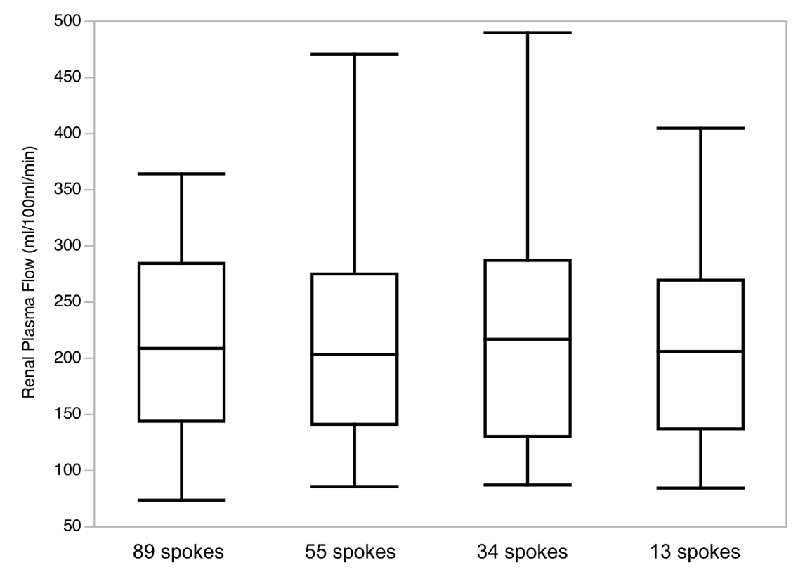
Using the reconstruction with the highest temporal resolution (13 spokes), mean rPF was 216±93ml/100ml/min. This is comparable to previously reported values for DCE-MRI derived rPF values. The analysis of the datasets with lower temporal resolution yielded similar rPF values with 219±97ml/100ml/min for 34 spokes; 217±96ml/100ml/min for 54 spokes and 209±80ml/100ml/min for 89 spokes (Figures 1-3). No statistically significant differences between the groups were found (p>0.09 for all pairs), while limits of agreements were narrower in the comparison of the groups with lower temporal resolutions.Discussion
The continuously acquired radial, golden-angle stack-of-stars sequence allowed an intra-individual comparison of DCE MRI perfusion datasets with different temporal resolution using a single contrast injection. Despite an up to 7-fold difference in temporal resolution, the deconvolution-approach-based quantitative analysis resulted in similar rPF values for all datasets. This is of interest for clinical routine, in which the trade-off between temporal and spatial resolution in DCE-MRI protocols is a typical dilemma. An increase in temporal resolution does not seem to result in relevant changes of rPF parameters. Previous studies suggested a temporal resolution of 4s in renal perfusion imaging5.Conclusion
DCE MRI for quantitative renal perfusion assessment performed with temporal resolutions between 1.5s and 10.1s results in comparable rPF values. Our data suggests that it might be reasonable to invest in spatial resolution or SNR/image quality rather than high temporal resolutionAcknowledgements
No acknowledgement found.References
1. Vallee JP, Lazeyras F, Khan HG, Terrier F. Absolute renal blood flow quantification by dynamic MRI and Gd-DTPA. Eur Radiol 2000;10(8):1245-52.
2. Mendichovszky I, Pedersen M, Frokiaer J, et al. How accurate is dynamic contrast enhanced MRI in the assessment of renal glomerular filtration rate? A critical appraisal. J Magn Reson Imaging 2008;27(4):925-31.
3. Riffel P, Zöllner FG, Budjan J, et al. "One-Stop Shop": Free-Breathing Dynamic Contrast-Enhanced Magnetic Resonance Imaging of the Kidney Using Iterative Reconstruction and Continuous Golden-Angle Radial Sampling. Invest Radiol 2016;51(11):714-9.
4. Zöllner FG, Daab M, Sourbron SP, Schad LR, Schoenberg SO, Weisser G. An open source software for analysis of dynamic contrast enhanced magnetic resonance images: UMMPerfusion revisited. BMC Med Imaging 2016;16:7.
5. Michaely HJ, Sourbron SP, Buettner C, Lodemann KP, Reiser MF, Schoenberg SO. Temporal constraints in renal perfusion imaging with a 2-compartment model. Invest Radiol 2008;43(2):120-8.
Figures
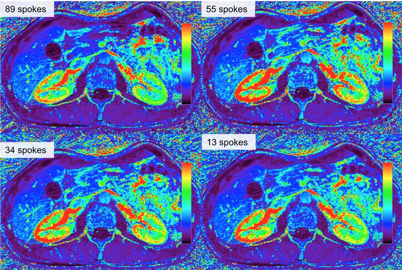
Color encoded maps for plasma flow of 4 datasets with different temporal resolutions in a single patient. Similar renal plasma flow values can be seen in all datasets.