3264
Time-Resolved Contrast-Enhanced MR Angiography with Single-Echo Dixon Background Suppression1Radiology, Mayo Clinic, Rochester, MN, United States
Synopsis
Contrast-enhanced MR angiography (CE-MRA) typically relies on a time-subtraction technique to suppress the background signal and emphasize the contrast-enhanced blood signal. However, Dixon-based background suppression has been reported to reduce motion sensitivity and improve signal-to-noise ratio in CE-MRA studies. Dual-echo Dixon techniques suffer a time penalty from acquiring an image at a second echo time, but single-echo Dixon techniques require an image at only one echo time and can reduce the time penalty. Here, time-resolved 3D single-echo Dixon CE-MRA at 3.0T with image update times of under 5 seconds is reported with results in the calves, hands, and brain.
Purpose
Contrast-enhanced MR angiography (CE-MRA) typically relies on a time-subtraction technique to suppress the background signal and emphasize the contrast-enhanced blood signal. Recently, however, Dixon-based background suppression has been reported to reduce motion sensitivity1 and improve signal-to-noise ratio (SNR)2 in CE-MRA studies. Traditional Dixon techniques use images at two different echo times to reconstruct separate images of water signal and fat signal in addition to the nuisance parameters associated with system imperfections. However, the increased acquisition requirements of the dual-echo Dixon technique compared to the time-subtraction technique (two echoes vs. one echo) reduce the temporal resolution of the dataset due to TR lengthening. Single-echo Dixon techniques3-7 require only a single image at a judiciously chosen echo time and assume that system imperfections are known a priori or may be otherwise estimated. Any lengthening of scan time compared to a subtraction exam (which has no echo time restrictions) is usually minor, and thus single-echo Dixon exams can provide scan times and temporal resolution comparable to time-subtraction exams while retaining an SNR improvement2. While the feasibility of time-resolved CE-MRA with dual-echo Dixon has been shown at 1.5T8, and dynamic single-echo Dixon imaging has been shown with temporal resolutions on the order of 30 seconds9, high temporal resolution time-resolved single-echo Dixon CE-MRA has not been reported. The purpose of this work is to report on time-resolved 3D single-echo Dixon CE-MRA at 3.0T with image update times of under 5 seconds.Theory
The phase-constrained signal equation for a spoiled gradient echo acquisition at echo time $$$t$$$ is shown in Eq. 1, where $$$\theta(t)$$$ is the chemical shift-induced phase, $$$\phi(t)=\gamma \Delta B_0 t$$$ is the phase due to magnetic field inhomogeneity, $$$\phi_0$$$ is the shared initial phase of $$$W$$$ and $$$F$$$, the real-valued water and fat signals, and $$$N(t)$$$ is zero-mean Gaussian noise. By constraining $$$W$$$ and $$$F$$$ to be real with shared initial phase, $$$\phi_0$$$, there are four real-valued unknown quantities10. These four unknown quantities can be estimated from two complex-valued measurements. Further, under the assumption that $$$\Delta B_0$$$ and $$$\phi_0$$$ are known or can be estimated4,11 (from a dual-echo calibration scan, for example), real-valued $$$W$$$ and $$$F$$$ can be reconstructed from a single complex measurement (Eq. 2).
$$G(t)=(W+Fe^{i \theta(t)})e^{i(\phi(t)+\phi_0)}+N(t)\hspace{1cm}\text{(Eq. 1)} $$
$$\begin{bmatrix}W\\F\end{bmatrix}=Re\{A^*A\}^{-1}Re\{A^*G\}\hspace{1cm}$$
$$\text{where }A=e^{i(\phi(t)+\phi_0)} \begin{bmatrix}1&e^{i\theta(t)}\end{bmatrix}\hspace{1cm}\text{(Eq. 2)} $$
Methods
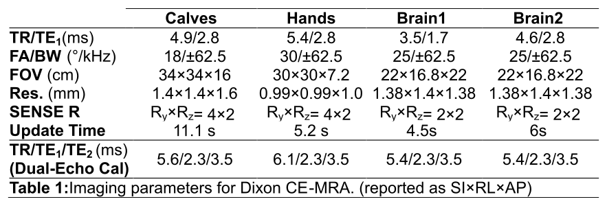
IRB-approved experiments were performed in healthy volunteers at 3.0T to evaluate the feasibility of highly accelerated time-resolved single-echo Dixon CE-MRA in a number of different anatomies. Studies were performed in the calves, hands, and brain with the scan parameters shown in Table 1. All studies used 10-12 mL of Gadobenate Dimeglumine (MultiHance, Bracco Diagnostics, Priceton, NJ) injected at 2 mL/s. Single-echo images were generated according to Eq. 2 using regularized maximum likelihood estimates of $$$\Delta B_0$$$ and $$$\phi_0$$$ obtained from dual-echo calibration images using a graph cuts-based optimization procedure.Results
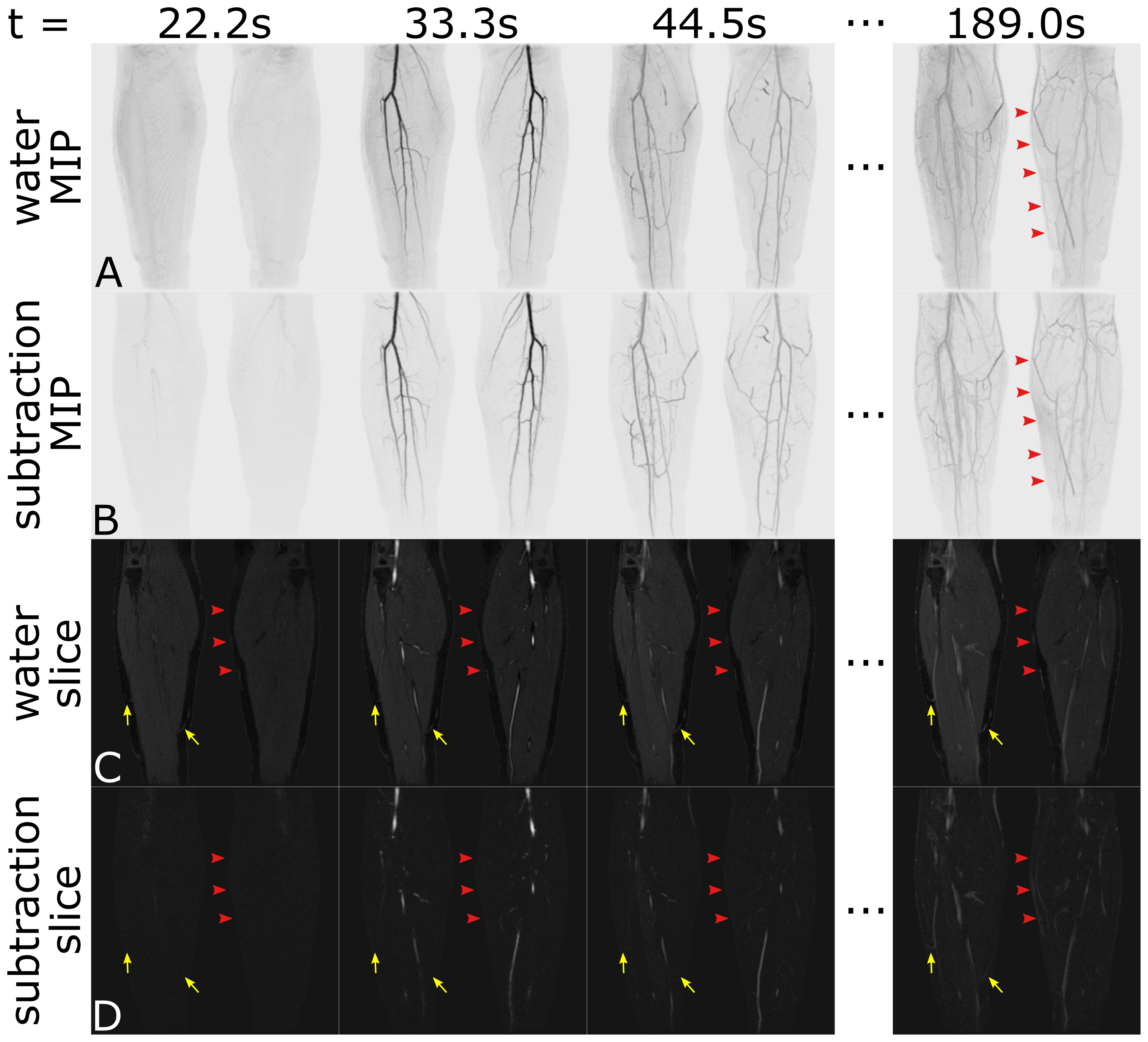
Time-resolved single-echo Dixon maximum intensity projections (MIPs) and images of the calves are shown in Figure 1A,C. Fat/water separation was successful for all time frames. Time subtraction MIPs and images are shown for comparison in Figure 1B,D. Red arrow-heads show motion-induced errors in the subtraction images that are not present in the Dixon images. Yellow arrows show improved depiction of small superficial subcutaneous veins within the fat in the Dixon images.
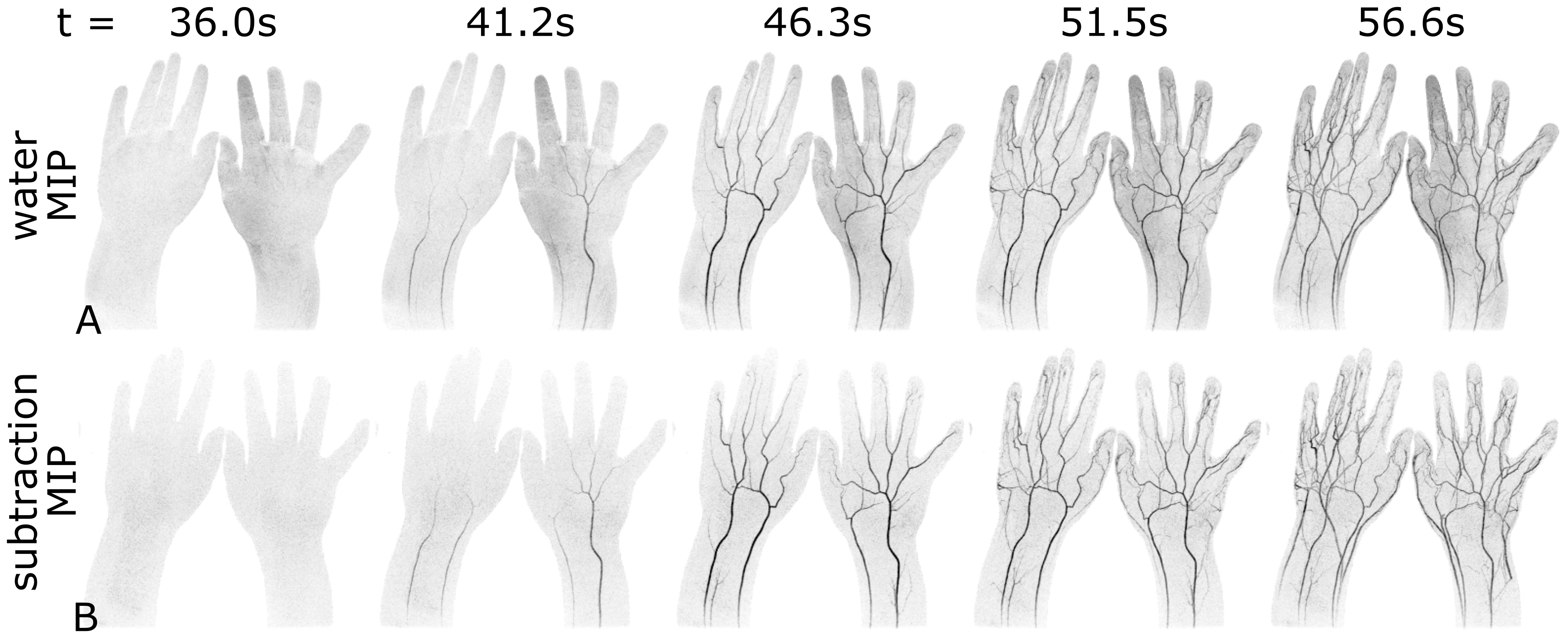
Figure 2 shows MIP results from a bilateral hand study with an image update time of 5.2s. Single-echo Dixon results (A) show excellent depiction of the vasculature, as do time-subtraction images (B).
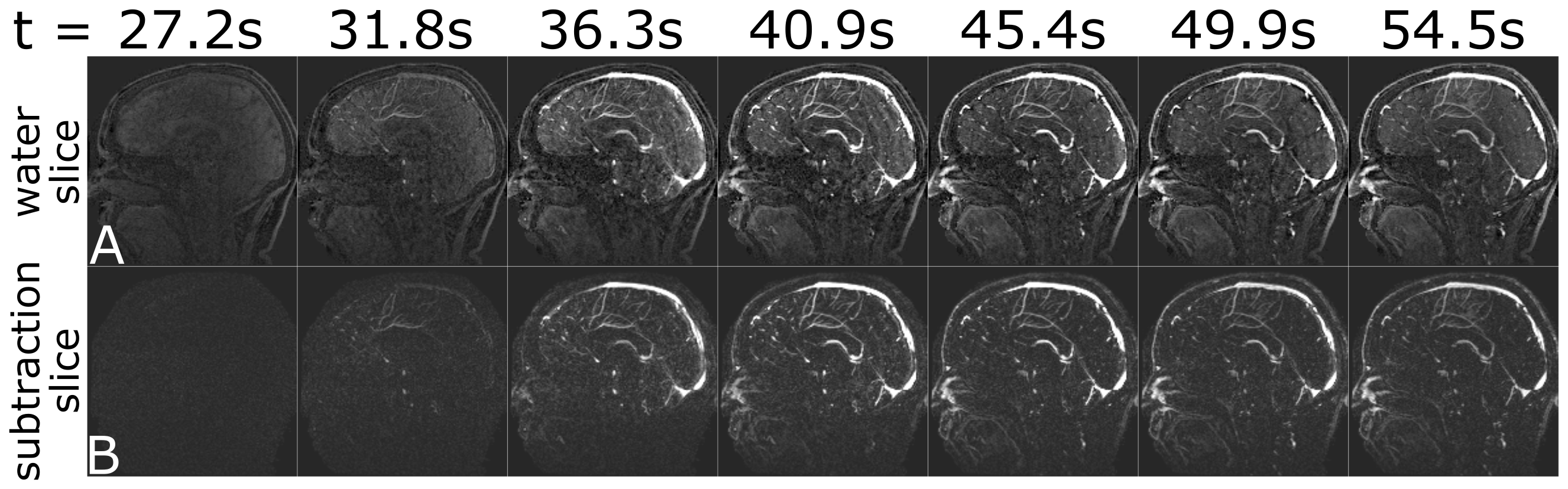
Brain images with an image update time of 4.5s are shown in Figure 3. Single-echo water images (A) include background water signal in addition to the CE blood signal, providing contextual anatomy.
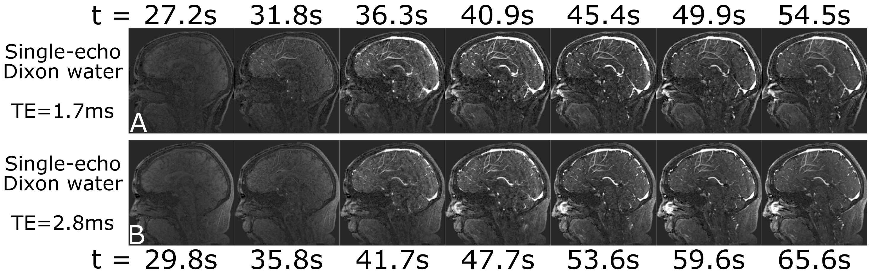
Single-echo Dixon has been shown to provide near optimal SNR in the water images at echo times that provide ±π/2 shift between the water and fat signal2, corresponding to TE=1.7ms and TE=2.8ms at 3.0T.
Figure 4 shows image results from studies in a single volunteer using these two echo times. Note the excellent fat suppression in both series, and reduced image update time for the shorter TE (which allows for a shorter TR).
Discussion
Time-resolved single-echo Dixon CE-MRA has been shown with highly accelerated imaging of the calves, hands, and brain. The images compare well with the time-subtraction results and the residual background signal is relatively benign. By reducing the echo time to provide a -π/2 chemical shift phase, image update time of <5s are possible in the brain. Future work will investigate improved image quality through advanced reconstructions, theoretical analysis of accelerated Dixon SNR, and applications to even more anatomies.Acknowledgements
Funded by: NIH EB000212, NIH RR018898, and DOD W81 XWH-15-1-0341References
1. Leiner T. Eur Radiol 2013;23:2228.
2. Stinson EG. MRM 2015;74:81.
3. Ma J. JMRI 2008;27:881.
4. Xiang Q-S. ISMRM 2001. #789.
5. Eggers H. ISMRM 2012. #1199.
6. Paltiel Z. SMRM 1985. #172.
7. Patrick J. SMRM 1985. #174.
8. Kouwenhoven M. ISMRM 2016. #886.
9. Yu H. MRM 2006;55:413.
10. Bydder M. MRI 2011;29:216.
11. Stinson EG. ISMRM 2016. #2673.
Figures