3259
QISS FISS (Quiescent-Interval Slice-Selective Fast Interrupted Steady-State): A Best-of-Both-Worlds Solution to Nonenhanced MR Angiography at 3 TeslaRobert R. Edelman1,2, Shivraman Giri3, and Ioannis Koktzoglou4,5
1Radiology, NorthShore University HealthSystem, Evanston, IL, United States, 2Radiology, Northwestern University, Chicago, IL, United States, 3Siemens HealthCare, 4Radiology, NorthShore University HealthSystem, 5Radiology, University of Chicago
Synopsis
QISS is a robust method for nonenhanced MRA that conventionally uses a TrueFISP readout. At 3 Tesla, use of TrueFISP is challenging due to high RF power deposition and sensitivity to off-resonance artifacts. In order to overcome these limitations, we implemented a QISS technique that incorporates a new type of readout that is a hybrid of TrueFISP and FLASH pulse sequences, which we call “Fast Interrupted Steady-State” (FISS). A pilot study was conducted to evaluate the potential benefits of QISS FISS at 3 Tesla for evaluation of the peripheral arteries and great vessels of the chest.
Purpose:
The QISS (quiescent-interval slice-selective) technique is a robust approach for nonenhanced MR angiography of the peripheral arteries (1), which also shows promise for evaluation of other vascular territories such as the great vessels of the chest and extracranial carotid arteries. Compared with 1.5 Tesla, QISS MRA at 3 Tesla benefits from an improved signal-to-noise ratio (SNR) and vessel-to-background contrast. However, QISS conventionally uses a balanced steady-state free-precession or TrueFISP readout, which poses significant challenges when applied at high field. For optimal results, the TrueFISP readout requires a large flip angle and short echo spacing, which at 3 Tesla may necessitate a large reduction in flip angle due to SAR limitations. At high field, TrueFISP images can be degraded by off-resonance and out-of-slice flow artifacts (e.g. banding and hot spots), which are often severe in the chest region. One option is to use a FLASH readout, which is less sensitive to off-resonance effects than TrueFISP and is not SAR-limited. However, a FLASH readout has other drawbacks including inferior SNR and saturation of slowly moving arterial spins. In order to overcome these challenges, we developed a novel pulse sequence which we call “FISS" (Fast Interrupted Steady-State). The FISS pulse sequence is essentially a hybrid of TrueFISP and FLASH pulse sequences that maintains the benefits of a TrueFISP readout while avoiding its limitations. In order to determine its potential benefits and limitations, we conducted a pilot study of QISS using a FISS readout for evaluation of the peripheral arteries and great vessels of the chest at 3 Tesla, and compared it with QISS using TrueFISP and FLASH readouts.Methods:
The QISS FISS pulse sequence consists of an initial in-plane saturation or inversion RF pulse, optional tracking saturation, quiescent interval, followed by the FISS readout. The sequence is ECG-gated such that the quiescent interval spans systole while the readout is timed to occur during slow diastolic flow. The FISS readout consists of repeated modules of the form: [+alpha/2,(-alpha,read,+alpha)n,-alpha/2,spoil], in which the imaging gradients are balanced aside from the final spoiler. The RF pulses applied after the readout function to tip the magnetization vector back along the longitudinal axis, thereby reducing saturation effects akin to a TrueFISP readout. For this study, n was set to a value of 1 with echo spacing of ~9-13 ms. This IRB-approved study was conducted on a 3 Tesla scanner (MAGNETOM Skyra-fit, Siemens Healthcare, Erlangen, Germany). A prototype QISS FISS sequence was compared with QISS TrueFISP and QISS FLASH. All sequences used a radial k-space trajectory. Typical slice thickness was 3-mm with in-plane resolution on the order of 1-mm. For peripheral MRA, 40 slices were acquired at each table station. For chest MRA, ~5-10 slices were acquired per breath-hold. Excitation flip angles ranged from 15 to 90 degrees. Because the echo spacing for both QISS FISS and QISS FLASH was longer than for a TrueFISP readout, 2 or 3 shots were typically acquired.Results:
QISS FISS showed markedly improved image quality, SNR and vessel-to-background contrast compared with a FLASH readout, plus a substantial reduction in image artifacts compared with a TrueFISP readout (Figure 1). Image quality for QISS FISS was much better than QISS FLASH for larger flip angles. For instance, using a flip angle of 89 degrees, QISS FISS provided excellent demonstration of through-plane arterial flow, whereas QISS FLASH showed severe image degradation and near complete suppression of arterial signal. Compared with QISS TrueFISP, QISS FISS showed more uniform vascular and background signal as well as less severe flow and off-resonance artifacts. The image quality of the QISS FISS technique was maintained through the entire image stack (e.g. 40 slices for peripheral vascular imaging), indicating that the method is stable away from the magnet isocenter.Discussion and Conclusion:
QISS FISS is a new imaging technique that provides major benefits for nonenhanced MRA at 3 Tesla. It largely avoids the image artifacts and SAR limitations that are encountered with a TrueFISP readout, while improving image quality and arterial enhancement compared with a FLASH readout. There are no steady-state signal contributions from vascular spins that have moved out of the slice, which avoids a common source of flow artifacts with TrueFISP (2). Initial results in the peripheral arteries and chest vessels appear promising. Further work will be directed towards sequence optimization and clinical validation. We also plan to explore the use of the FISS technique for cine imaging of the heart at 3 Tesla, where it has the potential to greatly improve image quality compared with cine TrueFISP.Acknowledgements
Study was funded by NIH grants R01 HL130093 and R21 HL126015.References
References. 1. Edelman et al. Magn Reson Med 2010; 63: 951. 2. Markl M, et al. Magn Reson Med. 2003;50:892.Figures
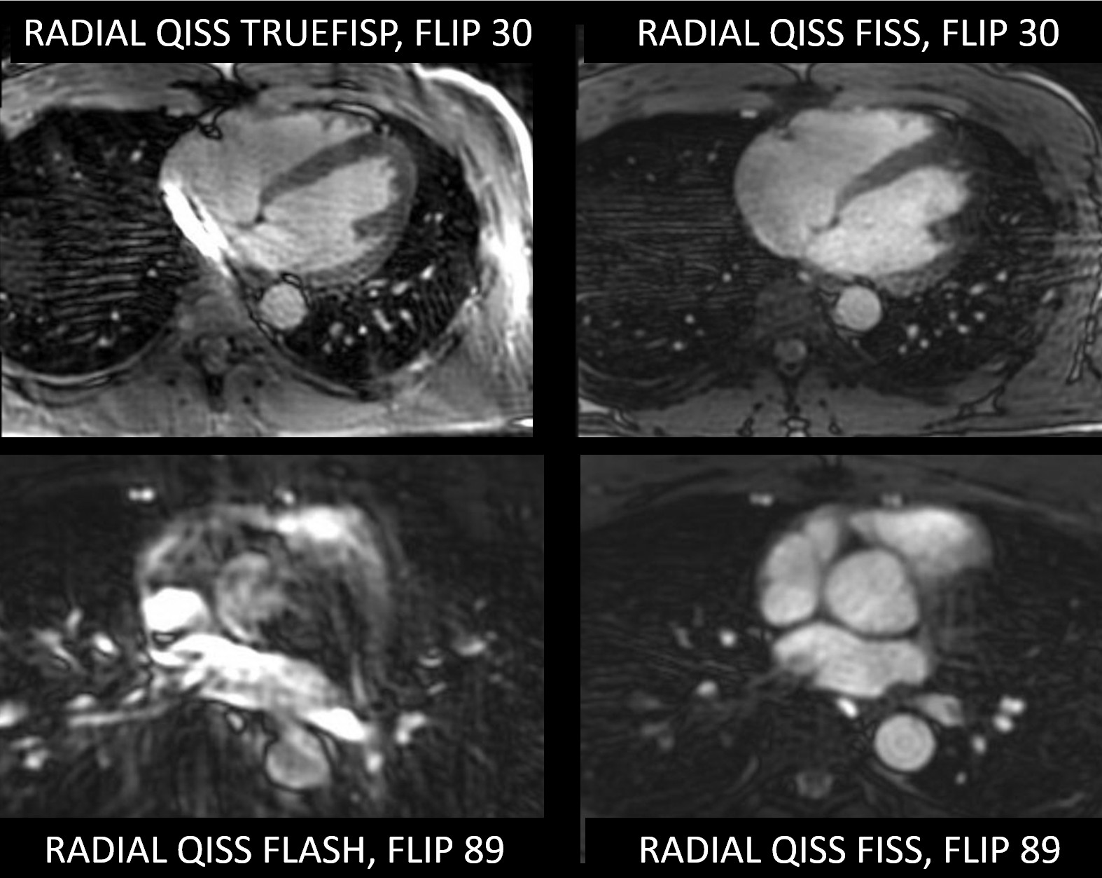
Figure 1. Comparison
of axial QISS images through the heart using various readouts at 3 Tesla. QISS TrueFISP with flip angle of 30 degrees
(upper left) shows a signal hotspot with severe anatomic distortion at the
medial margin of the heart, due to a combination of off-resonance and
out-of-slice flow effects. At the same
flip angle, QISS FISS (upper right) shows excellent flow contrast without
significant artifacts. At a flip angle
of 89 degrees, QISS FLASH (lower left) shows severe image degradation. At the same flip angle, QISS FISS (lower
right) shows excellent image quality.