3218
Comparison of SENSE, GRAPPA, SPIRiT and ESPIRiT for accelerated 4D flow MRI Imaging1Center for Biomedical Imaging Research, Department of Biomedical Engineering, School of Medicine, Tsinghua University, Beijing, People's Republic of China
Synopsis
Parallel imaging is a promising method to shorten the scanning time of 4D flow MRI. We applied SENSE, GRAPPA, SPIRiT and ESPIRiT in 4D flow imaging on six healthy volunteers and compared the accuracy of four algorithm. We found that ESPIRiT and SPIRiT showed the better velocity maps than SENSE and Grappa. The temporal fidelity of ESPIRIT can be the best among four methods, while SENSE and GRAPPA resulted in overestimates of peak flow. In conclusion, we validate the accuracy of four widely-used parallel imaging methods for the reconstruction of velocity map in 4D flow MR Imaging.
Purpose
Three-dimensional (3D) spatial encoding combined with three-directional velocity-encoded phase contrast information (4D flow MRI) has become an emerging method to measure the flow patterns and parameters in vessels [1]. However, one of the current limitations of 4D flow MRI is long scan times, in fact, a typical acquisition time for a standard 3D PC-MRI experiment for the aortic arch is around 20-30 min. This is significantly limiting the clinical application, while parallel imaging is one of methods promising to solve this problem for 4D flow MRI. Not much work has been done to evaluate the feasibility of accelerated 3D PC-MRI method with current parallel imaging methods. So the purpose of this study was to evaluate the accuracy of 4D flow imaging using different parallel imaging methods including SENSE [2], GRAPPA [3], SPIRiT [4] and ESPIRiT [5].Methods
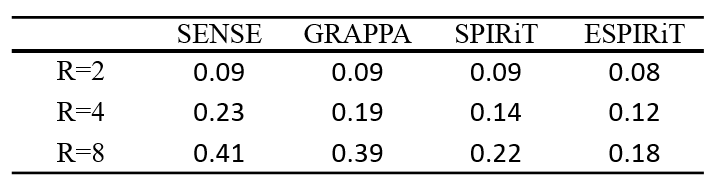
MRI Scan: The aortas of 6 healthy volunteers (mean age 25 years) were involved in this study. All images were fully-sampled on a 3.0 T whole-body MR scanner (Achieva, Philips Medical System, Best, The Netherlands) with a 32-channel cardiovascular coil. A sagittal slice across the ascending aorta, aortic arch, and descending aorta was imaged with 4D flow sequence. The relevant scanning parameters are: field of view (FOV) = 176 × 260 mm2 (FH/RL), slice thickness = 5 mm, spatial resolution = 2.6 × 2.6 mm2 (FH/RL), flip angle = 5°, repetition/echo time (TR/TE) = 6.2 ms/2.2 ms, number of cardiac phases = 32, temporal resolution is about 28 ms, and encoding velocity (VENC) = 150 cm/s. Undersampling strategy: We performed 3D retrospectively undersampled experiments in this study. The fully sampled 3D cine PC-MRI data were retrospectively undersampled with three net acceleration factors (i.e., R =2, 4, and 6) using ACS lines in central k-t space regions. The same sampling pattern was shared in four sets of PC-MRI (one reference set and three flow encoded sets). Comparison: We reconstructed the velocity maps using SENSE, GRAPPA, SPIRiT and ESPIRiT respectively. We performed analysis to evaluate the accuracy of different methods. To be specific, we defined the root-mean-square (RMSE) in a region of interest (ROI) for both the ascending aorta and descending aorta. To examine the temporal fidelity of different reconstruction methods, we also plot flow velocity over a cardiac cycle within an ROI in ascending aorta.Results
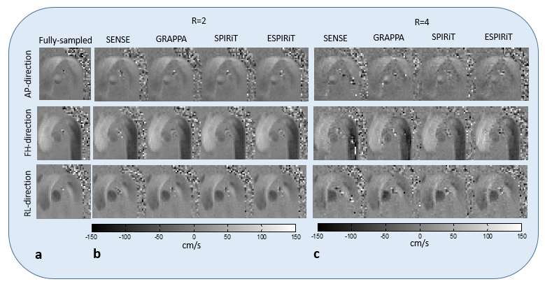
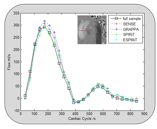
Undersampling 4D flow images with different acceleration factors were reconstructed through SENSE, GRAPPA, SPIRiT and ESPIRiT respectively are shown in Figure 1. ESPIRiT showed the best velocity maps among the four methods. The maps of four methods are similar at an acceleration factor of 2, when the acceleration factors come to 4, the velocity maps of ESPIRiT and SPIRiT are better than GRAPPA and SENSE. Actually, we could see some phase wrapping in the velocity maps of GRAPPA and SENSE for the descending aorta because of the over estimation of these two methods in some cases. Figure 2 showed reconstructed flow profiles along the FH-direction through the ascending aorta at the acceleration factor of 4, SENSE and GRAPPA resulted in overestimates of peak flow. In contrast, ESPIRiT captures the flow of the blood over the cardiac cycle best among the four methods.Discussion
The comparison of flow maps shown in Figure 1 and Table1 indicates that ESPIRiT and SPIRiT can keep accurate phase information at a reduction factor of 4, which is more accurate than GRAPPA and SENSE. According to Figure2, the temporal fidelity of ESPIRIT can be the best among four methods.Conclusion
We validate the accuracy of four widely-used parallel imaging methods (SENSE, GRAPPA, SPIRiT and ESPIRiT) for the reconstruction of velocity map in 4D flow MR Imaging.Acknowledgements
No acknowledgement found.References
[1] Markl M, Frydrychowicz A, Kozerke S, et al. 4D flow MRI[J]. Journal of Magnetic Resonance Imaging, 2012, 36(5): 1015-1036.
[2] Pruessmann K P, Weiger M, Scheidegger M B, et al. SENSE: sensitivity encoding for fast MRI[J]. Magnetic resonance in medicine, 1999, 42(5): 952-962.
[3] Griswold M A, Jakob P M, Heidemann R M, et al. Generalized autocalibrating partially parallel acquisitions (GRAPPA)[J]. Magnetic resonance in medicine, 2002, 47(6): 1202-1210.
[4] Lustig M, Pauly J M. SPIRiT: Iterative self-consistent parallel imaging reconstruction from arbitrary k-space[J]. Magnetic resonance in medicine, 2010, 64(2): 457-471.
[5] Uecker M, Lai P, Murphy M J, et al. ESPIRiT—an eigenvalue approach to autocalibrating parallel MRI: where SENSE meets GRAPPA[J]. Magnetic resonance in medicine, 2014, 71(3): 990-1001.
Figures