3192
Dynamic contrast enhanced 3D T1WI at upper abdomen using combination of parallel imaging and compressed sensing on a wide-bore 3T unit: Comparison of effects with Gd-DTPA and Gd-EOB-DTPAMotoyuki Katayama1, Takayuki Masui1, Kei Tsukamoto1, Mitsuteru Tsuchiya1, Yuki Hayashi1, Masako Sasaki1, Takahiro Yamada1, Yuji Iwadate2, Naoyuki Takei2, Kang Wang3, Kevin King4, and Harumi Sakahara5
1Radiology, Seirei Hamamatsu General Hospital, Hamamatsu, Japan, 2Global MR Applications and Workflow, GE Healthcare Japan, Hino, Japan, 3Global MR Applications and Workflow, GE Healthcare, Madison, WI, United States, 4Global MR Applications and Workflow, GE Healthcare, Waukesha, WI, United States, 5Radiology, Hamamatsu University School of Medicine, Hamamatsu, Japan
Synopsis
Consecutive four phases of dynamic contrast
enhanced 3D T1WI in the upper abdomen could be obtained during one breath-hold
in combined use of parallel imaging and compressed sensing at wide-bore 3T system.
The imaging protocols with Gd-DTPA and Gd-EOB-DTPA provided good image quality.
Although image contrasts with Gd-EOB-DTPA might be inferior to those with
Gd-DTPA, patterns of time intensity curves with study with dynamic contrast of
each protocol were similar to each other.
Introduction
MR imaging has played an important role in making a diagnosis of hepatobiliary and pancreatic diseases. Dynamic contrast three-dimensional (3D) T1-weighted imaging (T1WI) with Gadolinium (Gd) DTPA or Gd-EOB-DTPA has been one of the essential methods for the evaluation of the viability and characterization of the tumors. Because the injected amount of Gd-EOB-DTPA is half as much as that of Gd-DTPA, we might reduce the injection rate as half, 1.0-1.5mL/sec of Gd-EOB-DTPA to keep duration of data sampling of k-space. When scan time of conventional 3D T1WI is longer than 15 seconds, image quality might be degraded because of shortage of sampling data with Gd-EOB-DTPA. By using both parallel imaging and compressed sensing in combination, we can acquire the 3D images within 6 seconds at 3T wide-bore system.Purpose
The purpose of our study was to evaluate the dynamic contrast 3D T1WI with Gd-DTPA or Gd-EOBDTPA using the combination of parallel imaging and compressed sensing (CS-LAVA) with wide-bore 3T unit.Materials and Methods
Fifty-three patients, who underwent dynamic contrast enhanced MR images with 0.1mmol /kg of Gd-DTPA (n = 40) and 0.05mmol /kg of Gd-EOB-DTPA (n = 13) at wide-bore 3T unit (MR750W, GEHC), were included in this study. Dynamic contrast images using CS LAVA was obtained with MR smart-prep technique (Gd-DTPA; injection rates: 3 mL /sec, smart-prep triggering delay: 5 seconds, Gd-EOB-DTPA; injection rates: 1 mL/sec, smart-prep triggering delay: 5 seconds, respectively). The image reconstruction for ARC and CS was performed by applying CS iterative reconstruction followed by ARC data driven reconstruction in a serial manner1. Consecutive 4 phases (acquisition time: 6.1 seconds per phase) of images within one breath-held were obtained. Image quality of the dynamic phases of ach imaging was evaluated 5-point scale. The signal intensities of aorta, celiac artery, portal vein, right hepatic vein, liver parenchyma, pancreas, and spleen were measured on PACS system, respectively.Results
CS-LAVA images were acceptable; over all image quality; CS-LAVA with Gd DTPA: 4.2/ CS LAVA with Gd-EOB-DTPA: 4.0, lesion conspicuity: 4.6/4.5, blurring: 4.0/3.9, image distortion: 4.9/4.9, motion artifacts: 4.5/4.6, respectively. Graphs in Figure 1 and 2 indicate the image contrasts of each organ in subjective and objective analyses. In combined use of compressed sensing and parallel imaging for dynamic contrast study, the arterial phase can be obtained with both contrast agents. Although the image contrasts with Gd-EOB-DTPA were lower than those with Gd-DTPA, The patterns of the time-intensity contrast at each organ with each protocol were similar.Summary and conclusion
Dynamic contrast enhanced 3D T1WI with both Gd-DTPA and Gd-EOB-DTPA in combination of parallel imaging and compressed sensing at wide-bore 3T unit provide good image quality. The data in each phase could be obtained within a short period time as 6 seconds without viewsharing features causing possible contrast contamination. The provided pattern of the time-intensity contrast at each organ using Gd-DTPA and Gd-EOB-DTPA was similar.Acknowledgements
No acknowledgement found.References
King K, Xu D, Brau AC, Lai P, Beatty PJ, Marinelli L. A new combination ofcompressed sensing and data driven parallel imaging. Proceedings of theAnnual Meeting of ISMRM. Stockholm, 2010. (abstract 4881).Figures
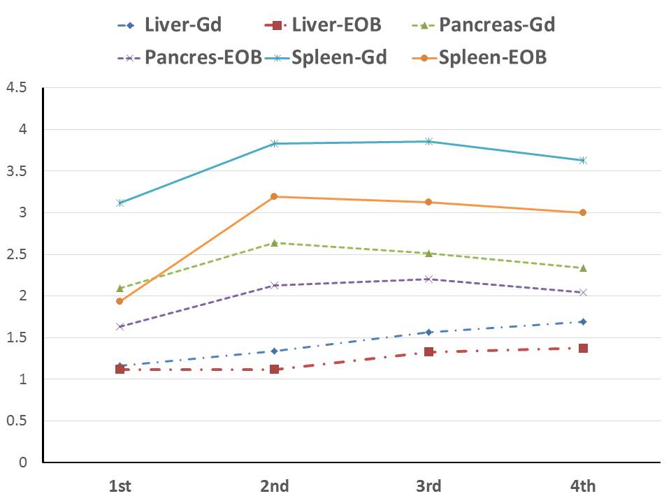
Quantitative analysis: The image contrasts
of lover parenchyma, pancreas, and spleen, respectively. The contrasts with
Gd-EOB-DTPA were inferior to those with Gd-DTPA. The pattern of the
time-intensity contrast at each organs with each protocols were similar.
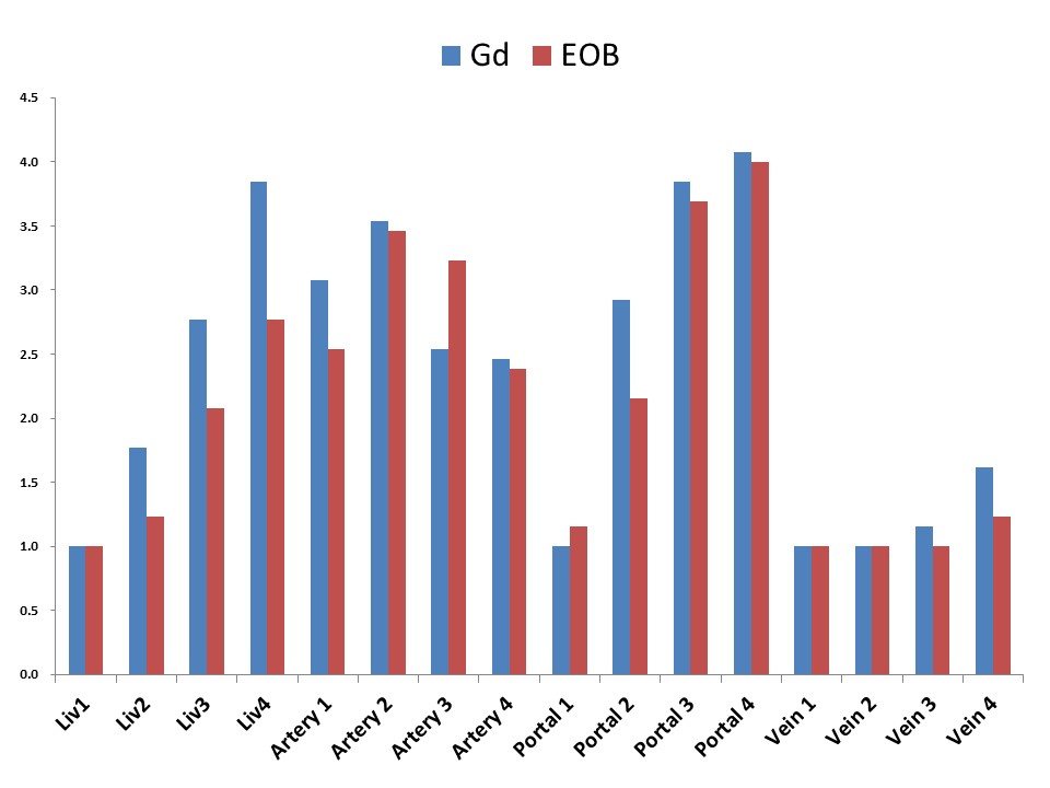
Qualitative analysis: The image contrasts of hepatic
parenchyma, pancreas, and spleen, respectively. The patterns of the signal intensities
at each structure with Gd-EOB-DTPA and Gd-DTPA were similar.
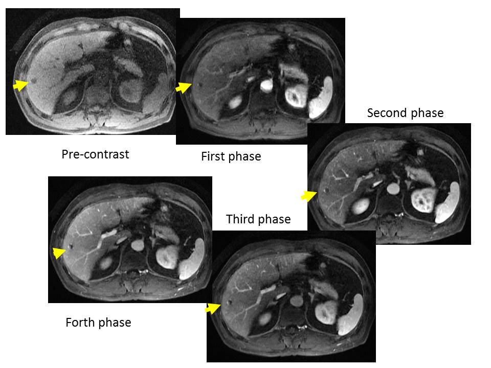
A male patient in his fifties with
hemangioma in segment 6 of his liver (arrow)
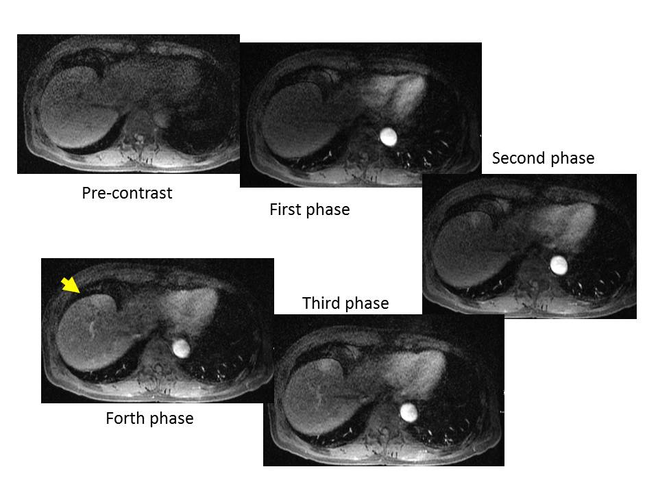
A male
patient in his seventies with hepatocellular carcinoma in segment 4 of his
liver (arrow