3175
Segmentation of Bone Marrow of Pelvis in Multi-parametric MRI (T1-w/ADC-map) of Metastatic Breast Cancer Patients1Quantitative MR Imaging and Spectroscopy Group, Research Center for Molecular and Cellular Imaging, Tehran University of Medical Sciences, Tehran, Iran, 2Department of Medical Physics and Biomedical Engineering, Tehran University of Medical Sciences, Tehran, Iran
Synopsis
To assess treatment response through quantitative analysis, in metastatic breast cancer patients, computer-aided segmentation of bone marrows is beneficial. We propose a semi-automatic segmentation method based on level-set and region growing techniques applied to T1-W images to facilitate extraction of apparent diffusion coefficient (ADC) features for the purpose of treatment response assessment from bone marrows of pelvic region. The results of applying the method on T1w/ADC-map of 10 patients shows a dice score of 81%, suggestive of high agreement of our proposed segmentation approach with expert’s opinion.
purpose
In advanced stages of breast cancer, bone marrow is one of the most probable regions for secondary tumors to form.1 Bone metastases impose serious complications for the patient, such as pain, suffering and poor quality of life. In this regard, accurate diagnosis, proper treatment planning, disease stage assessment and monitoring of treatment response are critical for improving the patient outcome. Quantitative approaches may pave the path towards reliable patient monitoring during the course of treatment, for which precise localization and delineation of whole lesional regions play are mandatory. Quantitative apparent diffusion coefficient (ADC) map derived from diffusion-weighted images (DWI) has high sensitivity to the changes in cell density, and therefore, can be used as non-invasive biomarker to show physiological changes in pathological tissues.2 Due to existence of different bone marrow types in bones, presence of metastases and bias field, bone marrow has a heterogeneous exhibition on MRI, which complicates the segmentation procedure. In this work, we aimed to implement an accurate segmentation technique for computer-aided extraction of bone marrow regions in presence of intensity inhomogeneity.Method
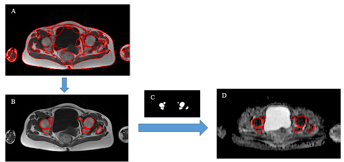
Whole-body T1-W and diffusion weighted (DW) images of 10 breast cancer patients with bone marrow metastases under treatment, were acquired on a 1.5T MR scanner (Avanto, Siemens). Due to low spatial resolution of ADC-maps, determination of lesions for quantitative assessment of treatment response is challenging. Therefore, lesion delineation should be performed on anatomical images (T1-w) and overlaid on corresponding ADC-maps to generate objective quantitative assessment. Here, we propose a semi-automated segmentation framework that can segment bone marrow in T1-Weighted images using a region-based level-set method that can handle the presence of intensity inhomogeneity through level-set evolution, followed by a region growing method. We applied the level-set algorithm proposed by Li et al3 on T1-W images, by assuming presence of slowly-varying intensity inhomogeneity within the images and trying to estimate and correct the inhomogeneity in several tissue clusters during the level-set evolution. The result of this step is a segmented image of whole regions, such as bone marrow, muscles and fat. For extracting bone marrow, we applied region growing technique by placing seed points on approximate locations of bone marrows, generating a mask of bone marrows to be overlaid on the corresponding registered ADC-maps of bone marrow tissues.Result
We applied our proposed algorithm (Fig. 1) on the images of the 10 patients and compared our results with manual delineations performed by an experienced radiologist using ImageJ software. According to table 1, the final assessment results show dice score~81%, specificity~99% and sensitivity~81%.
Discussion
Evaluation results indicate high agreement of the applied method with the expert’s opinion. We applied a semi-automatic technique to segment bone marrow in metastatic breast cancer patients, including two main steps: 1) a level-set method in presence of intensity inhomogeneity, followed by 2) region growing method for final segmentation of bone marrows. The proposed segmentation method facilitates the analysis of bone marrow lesions more accurately and reliably than manual selection of ROIs, which is time consuming, irreproducible in both intra- and inter-reader assessments, and prone to human errors. The method was applied on 10 patients and showed a Dice score of 81% indicating high segmentation performance.Conclusion
We proposed an efficient computer-aided diagnostic framework for reliable extraction of bone marrows from T1-W images for bone assessment.Acknowledgements
No acknowledgement found.References
1. Moulopoulos, Li A. Angel A., and V. Assilis Koutoulidis. Bone Marrow MRI. Springer Milan, (2015).
2. Padhani, Anwar R., Dow-Mu Koh, and David J. Collins. Whole-body diffusion-weighted MR imaging in cancer: current status and research directions. Radiology 261.3 (2011): 700-718.
3. Li, Chunming, et al. A level set method for image segmentation in the presence of intensity inhomogeneities with application to MRI. IEEE Transactions on Image Processing 20.7 (2011): 2007-2016.
Figures