3168
Motion Correction of Multi-b-value Diffusion-Weighted Images in the Kidney by Pyramidal Lucas-Kanade Registration1Academy for Advanced Interdisciplinary Studies, Peking University, Beijing, People's Republic of China, 2College of Engineering, Peking University, Beijing, People's Republic of China, 3Department of Radiology, Peking University First Hospital, Beijing, People's Republic of China
Synopsis
Intravoxel incoherent motion diffusion weighted imaging (IVIM-DWI) is a novel functional MRI technique which has been demonstrated with excellent diagnostic capability. Respiratory motion artifacts are the main source of error in the acquisition and quantification of parameters for multi-b-value DWI. This work develops a reliable approach to compensate for misalignments between multi-b-value images during free-breathing based on pyramidal Lucas-Kanade registration. Preliminary results show that our proposed approach can well correct motion-related artifacts misalignment. In addition, visual quality of the ADC, f, D and D* maps indicate that contours of kidney look better defined after registration.
Introduction
Diffusion-weighted magnetic resonance imaging (DWI) has been applied frequently for the noninvasive functional assessment of kidneys[1-3]. Its yielded quantitative parameter includes both apparent diffusion coefficient (ADC) representing diffusion of water molecules and perfusion fraction (Fp) indicating the microcirculation of blood in the capillary network[4]. However, DWI is sensitive to molecular displacement, bulk motion and physiologic motions such as respiration. What’s worse, in human subjects, respiratory motion may displace the kidneys by 5–10 mm, and this relative motion between images acquired at different time-points may introduce substantial error into the estimation of functional parameters[5, 6].
In this study, we propose a pyramidal Lucas-Kanade based image registration method to address motion-related artifacts during free-breathing in multi-b-value acquisitions.
Method
Free breathing diffusion datasets of ten subjects were performed at a 3-Tesla whole-body MR imaging unit (Discovery MR750; GE Medical Systems, Waukesha, WI) equipped with a 32-channel phased-array coil. The IVIM-DWI was acquired in two condition (respiratory triggered and free-breathing state) using a diffusion weighted single shot EPI fat saturated sequence with TR/TE 3300/57ms, matrix 128×128, 10 coronal slices, thickness 5mm, slice gap 2mm, GRAPPA factor 3, scan time 180s and ten b-values = 0,10,20,50,100,180,300,420,550, and 700s/mm2. The IVIM parameters derived before and after registration were compared for method evaluation.
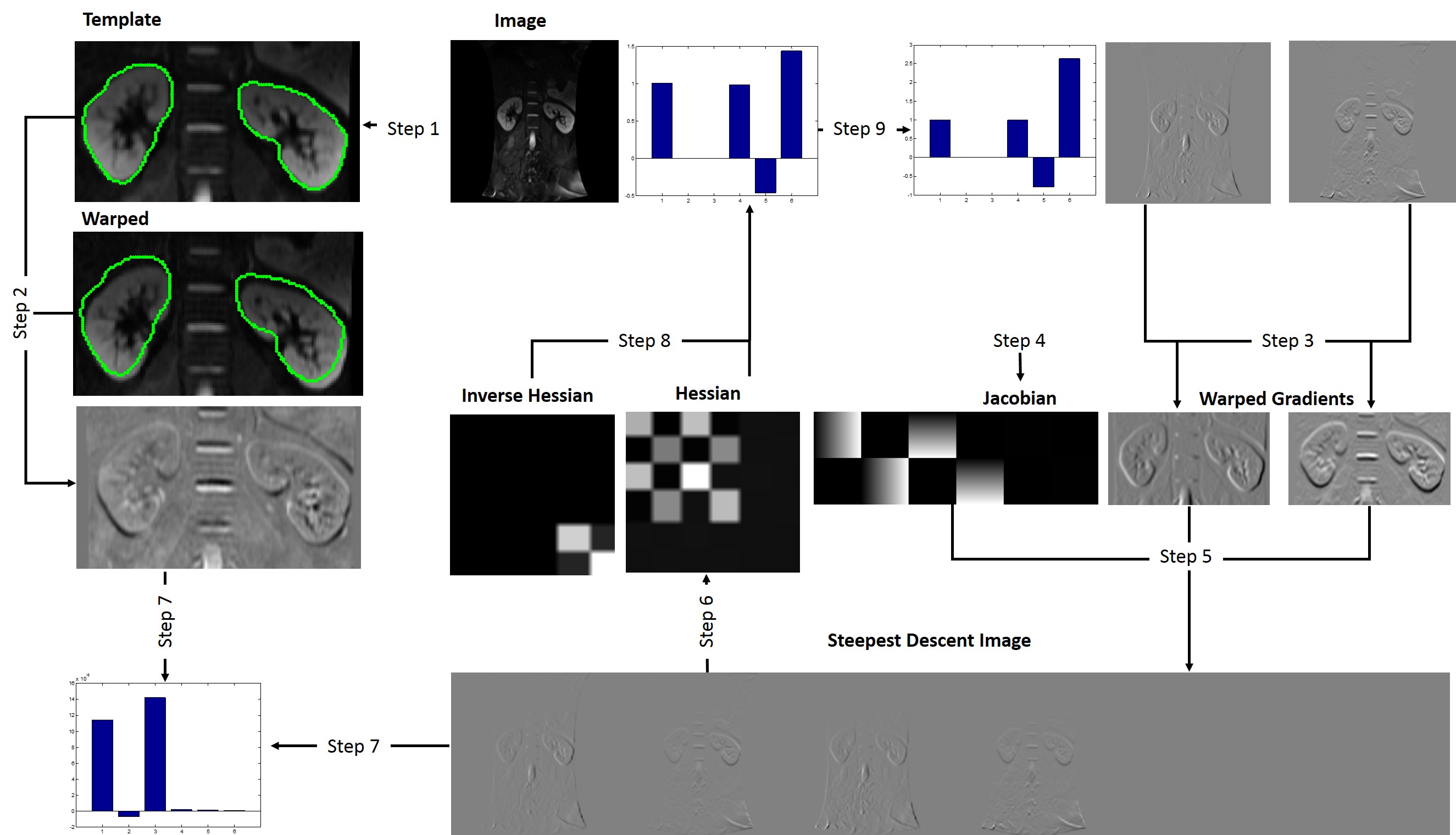
The main steps are illustrated in Figure 1 and described as follows. The proposed fully automatic pipeline consists of nine main steps. Firstly, the image I is warped with the current estimate of the warp in Step 1 and the result subtracted from the template in Step 2 to yield the error image. The gradient of I is warped in Step 3, the Jacobian is computed in Step 4, and the two combined in Step 5 to give the steepest descent images. In Step 6 the Hessian is computed from the steepest descent images. In Step 7 the steepest descent parameter updates are computed by dot producting the error image with the steepest descent images. In Step 8 the Hessian is inverted and multiplied by the steepest descent parameter updates to get the final parameter updates which are then added to the parameters in Step 9. The pyramidal structure in this study has 3 layers.
Result
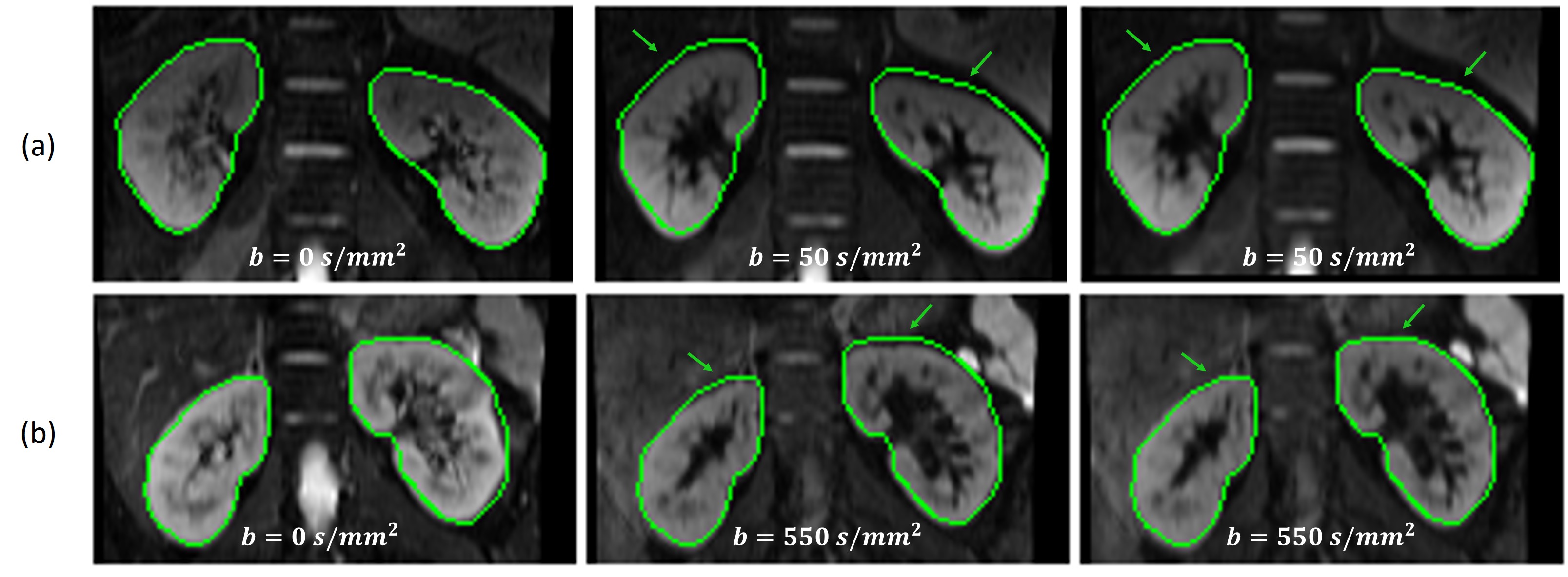
Figure 2 shows the registration results of the proposed motion correction method on images with different b-value. The green lines represent the edges of kidney in b0 image. Before registration, the kidney and the contour do not match. However, post-registered images indicate that our proposed method align the different b-value images to b0 image in the edge of kidney well.
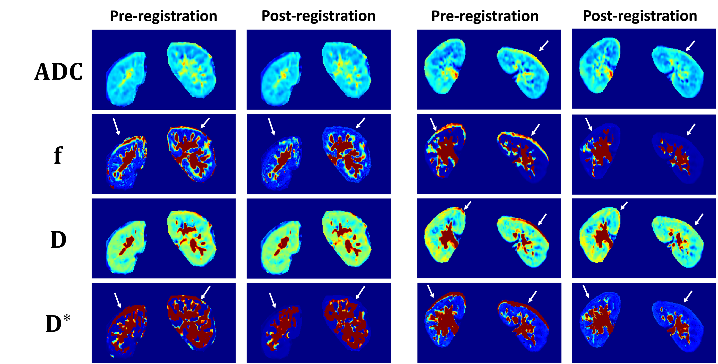
Figure 3 provides the comparison of the D, D*, f and ADC mapping before and after registration with our proposed method. Visual inspection suggest that the image registration scenarios improve the visual quality of these maps: contours of kidney are better defined and the number of voxels for which the fitting fails decreases.
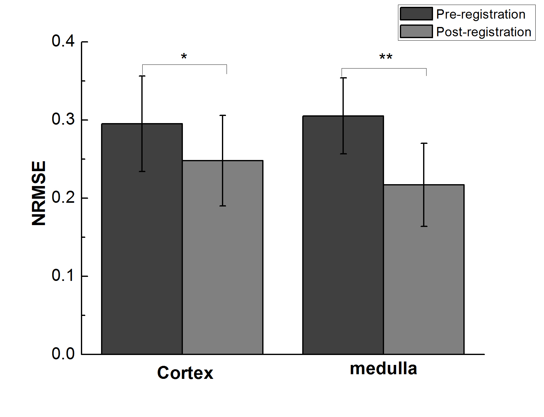
As shown in Figure 4, NRMSE was significantly lower after registration in both cortex (0.30±0.06 vs 0.25±0.06, P < 0.05) and medulla (0.31±0.05 vs 0.22±0.05, P < 0.001) of free-breathing measurements.
Discussion
In this study, we have proposed an image registration pipeline using pyramidal Lucas-Kanade to reduce artifacts caused by motion during free-breathing acquisition of DW images at multi-b-values.
The preliminary results demonstrate the feasibility of our method for aligning internal organs. The proposed method greatly increases the precision of IVIM fitting accuracy and quantity parameters estimation.
Its potential benefit in the assessment of abnormal kidney function will be explored in the future.
Acknowledgements
No acknowledgement found.References
1. Mike, N., M.F. Reiser, and S.P. Sourbron, Diffusion and perfusion of the kidney. European Journal of Radiology, 2010. 76(76): p. 337-47.
2. hoeny, H.C. and D.K. Frederik, Diffusion-weighted MR imaging of native and transplanted kidneys. Radiology, 2011. 259(259): p. 25-38.
3. Wittsack, H.-J., et al., Statistical evaluation of diffusion-weighted imaging of the human kidney. Magnetic Resonance in Medicine Official Journal of the Society of Magnetic Resonance in Medicine, 2010. 64(2): p. 616-22.
4. Mazaheri, Y., et al., Motion correction of multi-b-value diffusion-weighted imaging in the liver. Academic radiology, 2012. 19(12): p. 1573-1580.
5. Ms, C.C.C., et al., Performance of an efficient image-registration algorithm in processing MR renography data. Journal of Magnetic Resonance Imaging, 2015. 43(2): p. 391-397.
6. Siva, S., et al., An analysis of respiratory induced kidney motion on four-dimensional computed tomography and its implications for stereotactic kidney radiotherapy. Radiation Oncology, 2012. 8(11): p. 508-509.
Figures