3165
Role of diffusion-weighted imaging in distinguishing thoracoabdominal neuroblastic tumors of various histological types and differentiation grades1Department of Radiology, Beijing Children's Hospital, Beijing, People's Republic of China, 2Department of Pathology, Beijing Children's Hospital, Beijing, People's Republic of China
Synopsis
Purpose: To evaluate whether DWI allow discrimination of thoracoabdominal neuroblastic tumors of various histological types and differentiation grades. Materials and Methods: The DWI scans of the thoracoabdominal neuroblastic tumors in twenty-five children were retrospectively evaluated. DWI was performed with 2 b values of 0 and 800s/mm2 on a 3.0T MR scanner. Results: In the 25 cases, ganglioneuroma (GN) was in 3 cases, ganglioneuroblastoma (GNB) -Intermixed in 4, GNB-Nodular in 3 and neuroblastoma (NB) in 15. The ADC values of the NBs were significantly lower than those of GNs/GNBs (P<.001). The ADC of GNB-Nodular/NB was significantly less than that of GN/GNB-Intermixed (p<0.0001). In GNB-Nodular and NB, the tumors with poorly differentiated and undifferentiated lesions (n=12) had significantly smaller ADC than those with differentiated composition (n=6) (P=.0012). Conclusion: ADC of DWI is highly valuable for discriminating thoracoabdominal neuroblastic tumors of different histological types and subtypes.
Purpose
Diffusion-weighted MR imaging (DWI) has demonstrated a great potential to help distinguish benign from malignant tumors in pediatric body[1,2]. The International Neuroblastoma Pathology Classification (INPC), established in 1999 and revised in 2003, described four categories of peripheral neuroblastic tumors (pNTs): neuroblastoma (NB); ganglioneuroblastoma (GNB), nodular; GNB, intermixed and ganglioneuroma (GN). Although conventional magnetic resonance (MR) imaging is very difficult to differentiate different types of pNTs, diffusion-weighted MR imaging (DWI) may show correlated results with histology, and seems to be of great value for differential diagnosis [3-5]. The purpose of this study is to evaluate whether DWI allow discrimination of thoracoabdominal neuroblastic tumors of various histological types and differentiation grades.
Materials and Methods
The institutional ethical approval was obtained for the retrospective study. Children with pNTs in thoracoabdominal body, who were examined by DWI before any treatment, were included in this study. We collected a total of 25 cases (11 girls, 14 boys) in our database from April 2011 to August 2016. DWI was performed with 2 b values of 0 and 800s/mm2 on a 3.0T MR scanner. The DWI scans of the thoracoabdominal neuroblastic tumors were evaluated retrospectively. ADC values of different classifications of tumors were compared with Independent Samples T Test.Results
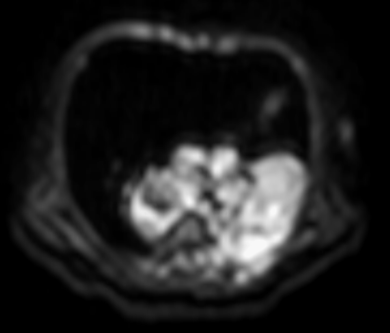
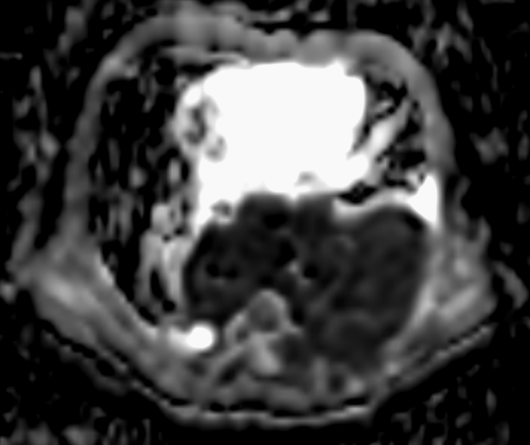
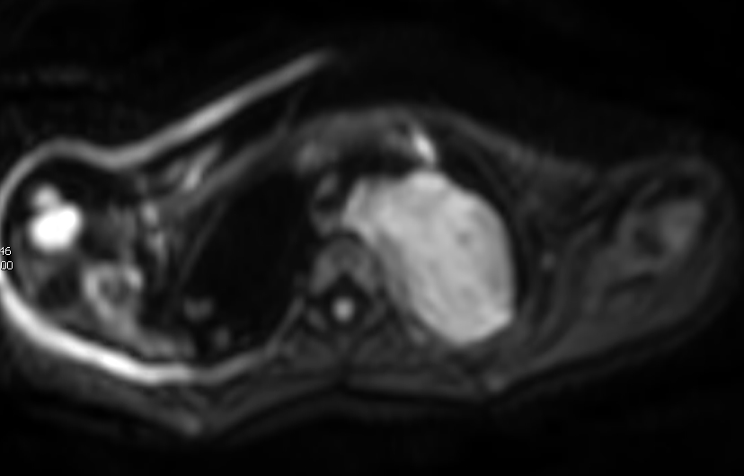
In the 25 patients with pNTs, GN was in 3 cases, GNB-Intermixed in 4, GNB-Nodular in 3 and NB in 15. The mean ADC of the 15 NBs was 0.82×10⁻³mm2/s (SD 0.17×10⁻³mm2/s, range 0.64–1.16×10⁻³mm2/s). And the mean ADC of the 3 GNs and 7 GNBs was 1.20×10⁻³mm2/s (SD 0.31×10⁻³mm2/s, range 0.62–1.49×10⁻³mm2/s). The NBs had significantly lower ADC values than the tumors of GN and GNB (P<.001). If the cases with GNB-Nodular and with NB are considered as a group (n=18), the cases with GN and with GNB-Intermixed as another group (n=7), there was a significant difference of the ADC between these two groups (p<0.0001). The mean ADC of the 3 GNBs-Nodular and 15 NBs was 0.83×10⁻³mm2/s (SD 0.22×10⁻³mm2/s, range 0.62–1.42×10⁻³mm2/s). And the mean ADC of the 3 GNs and 4 GNBs-Intermixed was 1.33×10⁻³mm2/s (SD 0.10×10⁻³mm2/s, range 1.17–1.49×10⁻³mm2/s). In 3 cases of GNB-Nodular, nodules of 2 cases were poorly differentiated and nodule of 1 case was differentiated. In 15 cases of NB, there were 8 cases with poorly differentiated neuroblastic component, 5 cases with differentiated and 2 cases with undifferentiated component. In the GNBs-Nodular and NBs, the tumors with poorly differentiated (n=10) and undifferentiated (n=2) neuroblastic component had significantly smaller ADC than those with differentiated composition (n=6) (P=.0012). The mean ADC of the former 12 pNTs was 0.75×10⁻³mm2/s (SD 0.15×10⁻³mm2/s, range 0.62–1.11×10⁻³mm2/s). And the mean ADC of the latter 6 pNTs was 1.01×10⁻³mm2/s (SD 0.24×10⁻³mm2/s, range 0.77–1.42×10⁻³mm2/s).Discussion and Conclusion
This study confirm that there is a significantly different ADC between NB and GN/GNB. And much more than this, we discover that the ADC of GNB-Nodular/NB is significantly less than that of GN/GNB-Intermixed, the GNB-Nodular/NB tumors with poorly differentiated and undifferentiated neuroblastic component have significantly lower ADC values than those with differentiated composition. All these strongly suggest that DWI has very high potential value for differentiation of thoracoabdominal neuroblastic tumors of various histological types and differentiation grades.Acknowledgements
This work was supported by Beijing Municipal Administration of Hospitals Incubating Program, Code:PX20171206.References
1 Kocaoglu M et al. Pediatric abdominal masses: diagnostic accuracy of diffusion weighted MRI. Magn Reson Imaging 2010; 28: 629-36.
2 Gawande RS et al. Role of diffusion-weighted imaging in differentiating benign and malignant pediatric abdominal tumors. Pediatr Radiol 2013;43:836-45.
3 Uhl M et al. MRI-diffusion imaging of neuroblastomas: first results and correlation to histology. Eur Radiol 2002;12:2335-8.
4 Gahr N et al. Diffusion-weighted MRI for differentiation of neuroblastoma and ganglioneuroblastoma/ganglioneuroma. Eur J Radiol 2011; 79:443-6.
5 Serin HI et al. Diffusion weighted imaging in differentiating malignant and benign neuroblastic tumors. Jpn J Radiol. 2016 ;34:620-4.
Figures