3162
Accelerated 2D Cine MRI Featuring Compressed Sensing and ECG-triggered Retro-gating1Siemens Healthcare GmbH, Erlangen, Germany
Synopsis
We present the combination of ECG-triggered retrospective gating and compressed sensing for segmented 2D Cine imaging at high spatiotemporal resolution. This enables capturing the complete cardiac cycle in segmented acquisitions, while significantly reducing the total acquisition time with compressed sensing and auto-calibration. The method was evaluated in 8 healthy volunteers and ventricular function parameters were compared to reference 2D Cine acquisitions featuring ECG-triggered retro-gating. Both methods resulted in comparable image quality and equivalent quantitative values for ventricular function parameters.
Introduction
Cardiac MR is the gold standard for the quantitative assessment of cardiac function. For this purpose, typically a stack of 2D slices covering the left ventricle is acquired in multiple breath-holds. In the recent years, compressed sensing has proven to significantly accelerate the data acquisition of 2D cardiac Cine imaging, while preserving image quality and diagnostic value1-3. However, up to now compressed sensing has mainly been shown in combination with real-time imaging and/or prospective ECG triggering, which does not cover the late diastole and can therefore lead to a slight underestimation of cardiac function. In this work, we address this limitation by combining ECG-triggered retro-gating and compressed sensing for segmented 2D Cine imaging at high spatiotemporal resolution. In-vivo experiments were performed in 8 volunteers and the method was compared to conventional ECG-triggered retro-gating.
Methods
ECG-triggered retro-gating allows adapting the length of the acquisition window to the duration of the heart-beat during continuous acquisition4,5. This enables capturing the complete cardiac cycle in segmented acquisitions. Typically, k-space interpolation4 or filtering5 is applied to retrospectively gate the acquired data to a reference heartbeat. This introduces an additional constraint to the data acquisition as sufficient k-space coverage must be ensured for conventional image reconstruction. Compressed sensing addresses this limitation with the ability to successful recover an image from incoherently sub-sampled data at high acceleration rates. In this context, retrospective gating can be translated into a binning procedure. Following a linear model, the acquired data of each heart beat is distributed to a constant number of phases, i.e., time frames. This binning procedure can be easily incorporated into the compressed sensing reconstruction and combined with auto-calibration6, which avoids the need to acquire additional calibration data.A prototype sequence and reconstruction implementing the proposed method were fully integrated in a $$$1.5$$$T clinical MR scanner (MAGNETOM Aera, Siemens Healthcare, Erlangen, Germany). In-vivo experiments were performed in $$$8$$$ healthy volunteers ($$$2$$$ female, age $$$47 \pm 20$$$ years), and the proposed method was compared to conventional retro-gating ECG-triggering. Cine images in $$$3$$$ long-axis orientations and a short-axis stack covering the whole left ventricle were acquired. For both methods, the following parameters were matched: TR$$$=2.8$$$ ms, TE$$$=1.2$$$ ms, $$$\alpha=58^\circ$$$, FOV$$$=360\times 270$$$ mm$$$^2$$$, pixel size$$$=(1.9$$$ mm$$$)^2$$$, slice thickness$$$=8$$$ mm, temporal resolution $$$37$$$ ms and a receiver bandwidth of $$$890$$$ Hz/px. The conventional method was accelerated by a factor of $$$2$$$, and internal reference lines were utilized for the calculation of GRAPPA weights, which reduced the total acceleration factor to $$$1.6$$$. In the proposed method , the Cartesian data acquisition was incoherently sub-sampled by a factor of $$$5.6$$$ and the coil sensitivity maps were estimated using auto-calibration. Compressed sensing reconstruction was performed as described in Liu7 with regularization factor $$$0.0003$$$ and was terminated after $$$40$$$ iterations. For evaluation, we compared acquisition time and ventricular function (VF) parameters (ejection fraction, end-diastolic, and end-systolic volume) that were determined from the resulting images of both methods after manual segmentation of the left ventricle with syngo.via (Siemens Healthcare, Erlangen, Germany).
Results and Discussion
Data acquisition was successful in all volunteers. Figure 1 shows the reconstructed images of the end-systolic and end-diastolic phases exemplarily for one volunteer for both methods. While maintaining the same spatiotemporal resolution, the acquisition time per slice could be reduced from $$$8$$$ heartbeats, using the conventional approach, to $$$2$$$ heartbeats with the proposed method. The images of both approaches resulted in comparable image quality and equivalent quantitative assessment of VF (see Table 1). Alternatively, the speed up in acquisition time could be invested in improving the temporal and/or spatial resolution. Figure 2 provides an example for a scan with even higher temporal resolution ($$$11$$$ ms) compared to the conventional retro-gated approach ($$$37$$$ ms) while maintaining the same acquisition time.
Conclusion
Compressed sensing and ECG-triggered retro-gating successfully enabled capturing the complete cardiac cycle with highly-accelerated, segmented 2D cine imaging. The combination of compressed sensing and auto-calibration significantly reduced the acquisition time for the same spatiotemporal resolution while maintaining image quality and equivalent quantitative cardiac function parameters. The promising results in volunteers as well as the full integration of the proposed method in a standard clinical scanner enable a comprehensive evaluation in patients with different pathologies in the near future.Acknowledgements
References
1 Vincenti G, et al., Compressed Sensing Single–Breath-Hold CMR for Fast Quantification of LV Function, Volumes, and Mass, JACC 2009, 54(15):882-892
2 Feng L, et al., Highly-Accelerated Real-Time Cardiac Cine MRI Using k-t SPARSE-SENSE, MRM 2013, 70(1):64-74
3 Goebel J, et al., Compressed sensing cine imaging with high spatial or high temporal resolution for analysis of left ventricular function, JMRI 2016 44(2):366-374
4 Lenz GW, et al. Retrospective Cardiac Gating: A Review of Technical Aspects and Future Directions, MRI 1989, 7:445-455
5 Madore B, et al., Retrospectively gated cardiac cine imaging with temporal and spatial acceleration, MRI 2011, 29:457-469
6 Kellman P, et al. Adaptive sensitivity encoding incorporating temporal filtering (TSENSE). MRM 2001, 45:846–852.
7 Liu J, et al., Dynamic cardiac MRI reconstruction with weighted redundant Haar wavelets, ISMRM 2012, #4249
Figures
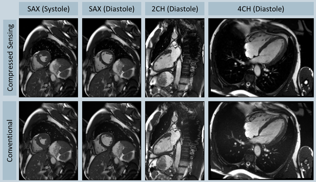
Figure 1: Comparison of 2D Cine images for one volunteer showing the short-axis (SAX) slices in diastole and systole, as well as two-chamber (2CH) and four-chamber (4CH) view for the proposed method (with compressed sensing) and the conventional approach.
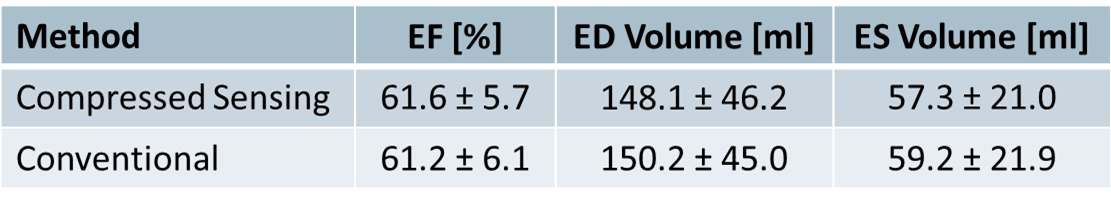
Table 1: Comparison of the quantitative results of the assessment of ventricular function for both methods.
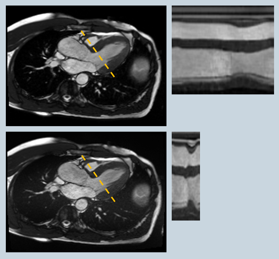
Figure 2: Comparison of 2D Cine images in three-chamber view featuring the same acquisition time. A temporal profile was plotted along the dashed line. While a temporal resolution of 37 ms can be achieved with conventional retro-gated 2D Cine imaging, the combination of compressed sensing and auto-calibration improved the temporal resolution to 11 ms.