3155
The utility of fetal MRI using real-time cine imaging for the functional assessment of congenital cardiovascular abnormality1Department of Radiology, National Cerebral and Cardiovascular Center, Suita, Osaka, Japan, 2Department of Perinatology and Gynecology, National Cerebral and Cardiovascular Center, Suita, Osaka, Japan
Synopsis
Volumetric analysis of the fetal heart by ultrasound (US) in congenital heart disease is often difficult due to its complex anatomy and US-specific artifacts. We implemented the real-time cine sequence without ECG triggering and the post-processing technique (PhyZiodynamics), which enabled noise reduction and interpolation based on motion coherence. Fetal MRI using real-time cine imaging allowed for detailed functional assessment in both ventricles and showed acceptable levels of correlation with both the prenatal and postnatal US findings, suggesting that this technique is a promising diagnostic tool for functional assessment of congenital cardiovascular abnormalities.
Introduction
The current gold standard in fetal cardiac imaging is the use of ultrasound (US). However, accurate volumetric analysis of both ventricles is often difficult in cases with congenital heart disease due to complex anatomy and US specific artifacts. Furthermore, US has the disadvantages of a limited acoustic window and observer dependency. Fetal MRI has recently become a viable tool for evaluating the fetal cardiovascular system. 1,2 One of the main issues in evaluating fetal cardiac function via cine imaging is gating the acquisition to the fetal ECG. Notably, the ECG triggering method for a fetus is not yet clinically available at all hospitals.Purpose
In this study, we performed fetal MRI using real-time cine imaging without ECG triggering and the post-processing technique (PhyZiodynamics), which enable noise reduction and interpolation based on motion coherence, and evaluated the feasibility of fetal MRI for functional assessment of the fetal heart for congenital heart disease.Methods
Prenatal cardiac MRIs on 18 pregnant women carrying fetuses with congenital heart disease were performed on a 1.5 T clinical machine (MAGNETOM Sonata, Siemens AG Healthcare Sector, Erlangen, Germany). Before cardiac MR examination, the fetal heart rate was measured via cardiotocography, and the detected heart rate was used as an R-R interval of simulated ECG in real-time cine imaging. Both the transverse and short-axis planes were acquired using a real-time multi-slice True-FISP cine sequence (TE/TR=1.3/2.6msec, flip angle 50°, FOV 200mm, matrix 192×154, slice thickness 4 mm) without ECG triggering of a fetal heartbeat. The length of real-time scanning was adjusted to two times the R-R interval (decided before MRI) using a simulated ECG. With use of the post-processing technique, which enabled noise reduction and interpolation based on motion coherence (PhyZiodynamics; Zio station 2 software, Ziosoft Inc., Japan), the effective temporal resolution was 37 msec. The left ventricular volume was analyzed from the short axis plane, and the right ventricular volume was analyzed from the transverse axis plane by two experienced observers via manual tracing of the endocardial border using Siemens Argus software. CMR findings were compared with prenatal and postnatal echocardiography.Results
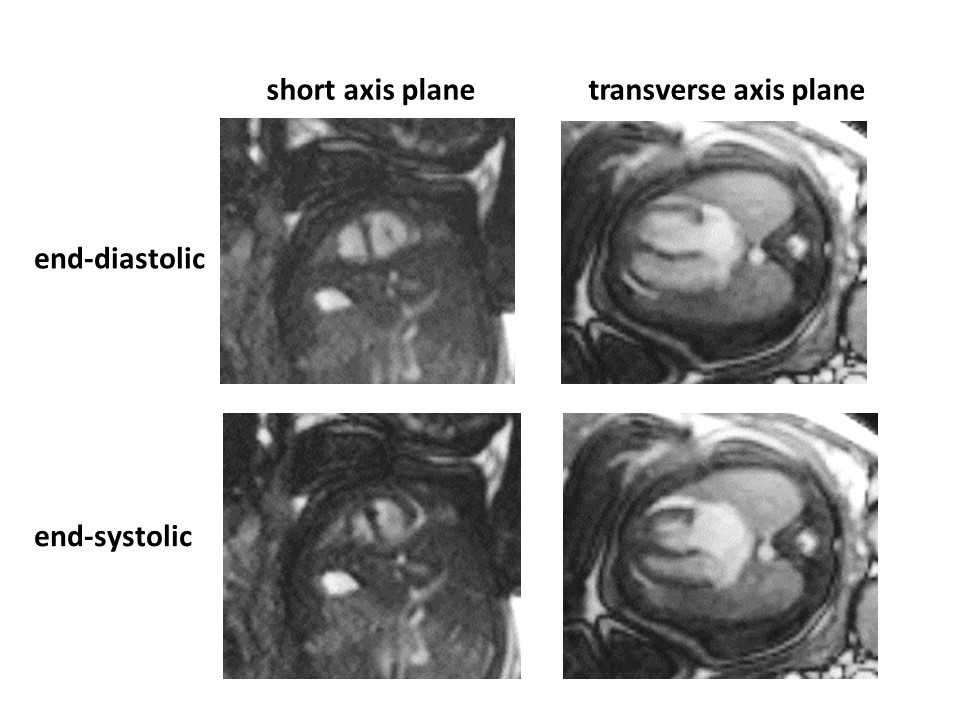
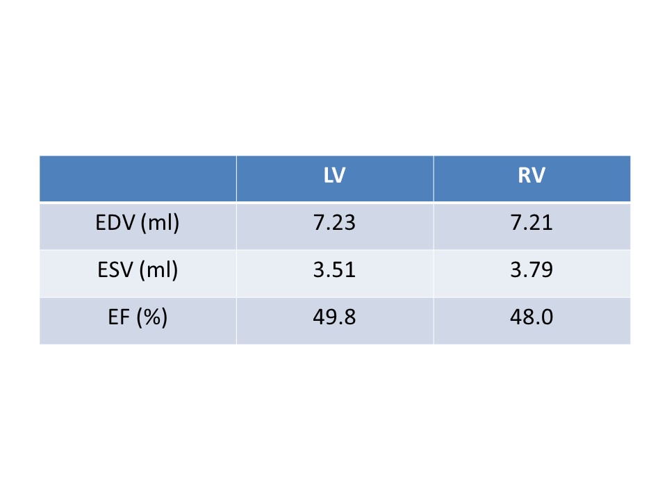
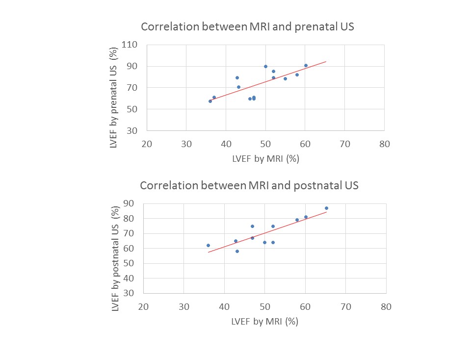
In all fetal images acquired from the real-time cine sequence, myocardial contraction of both ventricles was visualized well (Figure 1). Table 1 showed the mean end-diastolic volume (EDV), end-systolic volume (ESV), and ejection fraction (EF) via real-time cine imaging. LVEF based on fetal MRI and prenatal and postnatal US correlated well; prenatal US was r=0.73, and postnatal US was r=0.84 (Figure 2). LVEFs obtained by an MRI tended to be lower than those obtained with US. EDV on the fetal MRI and ventricular internal dimension in diastole (VDd) on the postnatal US also correlated well; LV was r=0.64, and RV was r=0.80 (Figure 3). Intra- and inter-observer variability for EDV, ESV, and EF in both ventricles were sufficiently small in real-time True-FISP cine imaging.Discussion
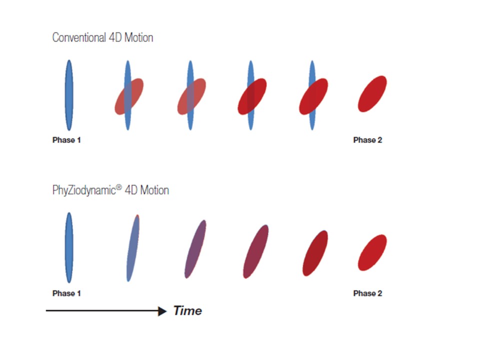
Our results demonstrated that real-time cine imaging without real ECG triggering allows detailed functional assessment in both ventricles and shows acceptable levels of correlation with both the prenatal and postnatal US findings. In this study, we used the novel post-processing technique PhyZiodynamics, which tracks the movement of individual voxels through space and time based on the non-rigid registration between phases (Figure 4). This voxel-to-voxel mapping of information enables the employment of additional algorithms that reduce noise, improve motion coherence, and measure function. 3 With use of this application, non-linear interpolation between registered voxels can generate increased phase data from the original cine data set. EFs were systematically lower when obtained with an MRI than with US; however, this might have been due to the different techniques employed. A previous study in a cardiac porcine model of known volume showed that cine MRI yielded the most precise volumetry by using methods similar to those used in our study, particularly for the right ventricle, which has a more complex anatomy. 4 Therefore, we believe that cine MRI has the potential to evaluate more accurate functional assessment compared with other modalities such as US.Conclusion
Fetal CMR using real-time cine imaging is a promising diagnostic tool for functional assessment of congenital cardiovascular abnormalities.Acknowledgements
No acknowledgement found.References
1. Dong SZ et al. Preliminary experience with cardiovascular magnetic resonance in evaluation of fetal cardiovascular anomalies. J Cardiovasc Magn Reson. 2013 May 21;15:40.
2. Manganaro L et al. Fetal MRI of the cardiovascular system: The role of steady-state free precession sequences for the evaluation of normal and pathological appearances. Radiol Med. 2009 Sep;114(6):852-70.
3. Brown HA PhyZiodynamics® A Revolutionary Approach for Post-Processed Noise Reduction, Motion Coherence, and Functional Analytics. 2010 Ziosoft, Inc.
4. Heusch A et al. Volumetric analysis of the right and left ventricle in a porcine heart model: Comparison of three-dimensional echocardiography, magnetic resonance imaging, and angiocardiography. Eur J Ultrasound. 1999 Jul;9(3):245-55.
Figures