3150
Myocardial Strain Imaging with Feature Tracking MRI in Patients with Cardiac Amyloidosis: Comparison with Normal Control Subjects1Radiology, Mayo Clinic, Rochester, MN, United States
Synopsis
Feature tracking (FT) myocardial strain analysis was performed in 87 patients with biopsy proven cardiac amyloidosis and in 64 control subjects using long and short axis ECG-triggered b-SSFP images and commercial software. Radial, circumferential, and longitudinal peak systolic strain values were all significantly different between amyloid patients and control subjects, suggesting that this technique is feasible in assessing patients with suspected cardiac amyloidosis.
Purpose
To compare myocardial strain values measured in patients with biopsy-proven cardiac amyloidosis with strain measurements in control subjects without significant cardiac disease using feature-tracking (FT) MRI.
Materials and Methods
Subjects: 87 patients were identified with biopsy-proven cardiac amyloidosis who had undergone clinically indicated cardiac MRI including short axis and long axis cine b-SSFP images. 64 subjects were identified to serve as controls who had cardiac MRI performed for clinical or research indications without imaging or clinical evidence of significant cardiac disease.
MRI: Cardiac MRI was performed on a 1.5T system (GE Twin Speed Excite) and included short axis and at least one long axis ECG-triggered cine b-SSFP acquisition with parameters including: TR/TE 3.4/1.2 ms, flip angle 50, slice thickness 8 mm, matrix 224x192, FOV 32 - 46 cm, 20 - 30 phases, 16 - 20 views per segment.
Analysis: Commercial software (Circle Cardiovascular Imaging) was used for FT strain analysis. End diastolic epicardial and endocardial borders were manually traced on short axis and long axis images with automated edge detection of remaining phases and strain analysis. Global radial and circumferential peak systolic strain values were recorded from short axis images as well as basal and apical values. Global peak systolic longitudinal strain was measured from long axis images. A 2-tailed Student's t-test was used to assess for significant differences between amyloid and control groups.
Results
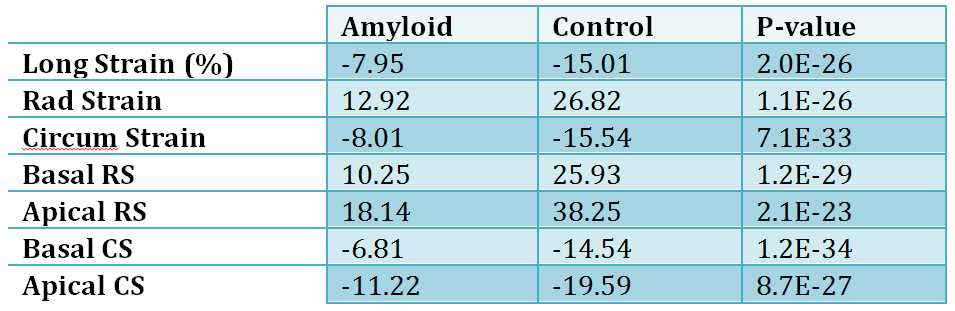
Amyloid patients consisted of 73 men and 14 women, average age 68 years, while the control subjects constituted 34 men and 30 women, average age 56 years. FT myocardial strain values and parametric images were successfully generated in all patients. There were significant differences in all strain categories measured, with p < 1x10-22 in all categories (Table 1, Fig. 1). Peak global radial, circumferential, and longitudinal strain values as well as basal and apical radial and circumferential strain values were lower in amyloid patients in comparison to control subjects, with several amyloid patients demonstrating more prominently reduced basal strain values with preserved apical strain (Fig. 2).Discussion
Myocardial strain analysis using FT MRI can be performed in most patients, with successful generation of parametric strain images dependent on relatively artifact-free cine b-SSFP images. Our results demonstrate highly significant differences between cardiac amyloid patients and control subjects in all strain parameters measured, suggesting that this is a feasible and potentially useful technique in assessing patients with suspected cardiac amyloidosis. Future investigations include assessment of the prognostic value of various strain measurements in amyloid patients, the utility of right ventricular strain measurements, and assessment of strain rate data.Acknowledgements
No acknowledgement found.References
No reference found.Figures