3143
Accelerated Cardiac Cine “Watermark” MRI provides Cardiac Function via Magnitude Cine and 2D Myocardial Strain via Spatially Modulated Phase1MRI Research Center, Auburn University, Auburn, AL, United States, 2Department of Engineering Science, University of Oxford, Oxford, United Kingdom, 3Sackler School of Graduate Biomedical Sciences, School of Medicine, Boston, MA, United States, 4Radiology and Imaging Sciences, National Institutes of Health, Bethesda, MD, United States, 5MR R&D, Siemens Healthcare, Malvern, PA
Synopsis
We
developed a parallel imaging accelerated Cine Watermark (CWM) sequence to
provide normal cine magnitude images plus phase image-only multi-directional
spatial encoding for quantitative cine strain -- while requiring no extra
operator effort. Spatial cosine
modulation post-processed by complex image sum/differencing produced separate
normal magnitude cine and unique phase-only spatial modulation for strain
calculation. In vivo human scans demonstrated good magnitude cine and phase-only
quantified displacement. Cardiac
strains were tracked by a novel non-linear least squares method combining
quadratic b-spline contours, nearest neighbor optimized phase tracking, and
physically-motivated regularizers. Cardiac left ventricular, short-axis,
circumferential strain results agreed with previous literature.
Purpose
Introduced over 25 years ago, myocardial tagging is installed on all manufacturers’ MRI scanners1-3. Despite extensive validation as the best method for clinical quantitative measure of myocardial wall deformation, tagging is limited to a research context due to the time-intensive analysis and need to acquire additional sequences. Cardiac MRI (CMR) sequences must provide maximum diagnostic information, and maximum patient throughput, with minimum operator effort and time (cost)4. We developed an accelerated CMR cine sequence with a “hidden” added capability to acquire cardiac strain data while requiring no extra operator effort. Called Cine Watermark (CWM), this sequence acquires normal cine magnitude images plus spatial-modulated tagging added only into the image phase (“watermark”) allowing quantitative cine strain calculation.Methods
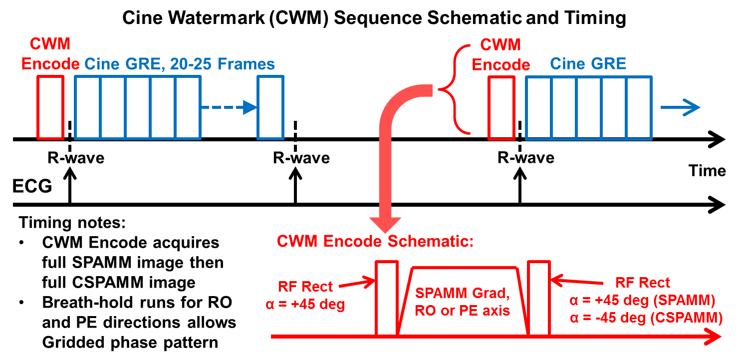
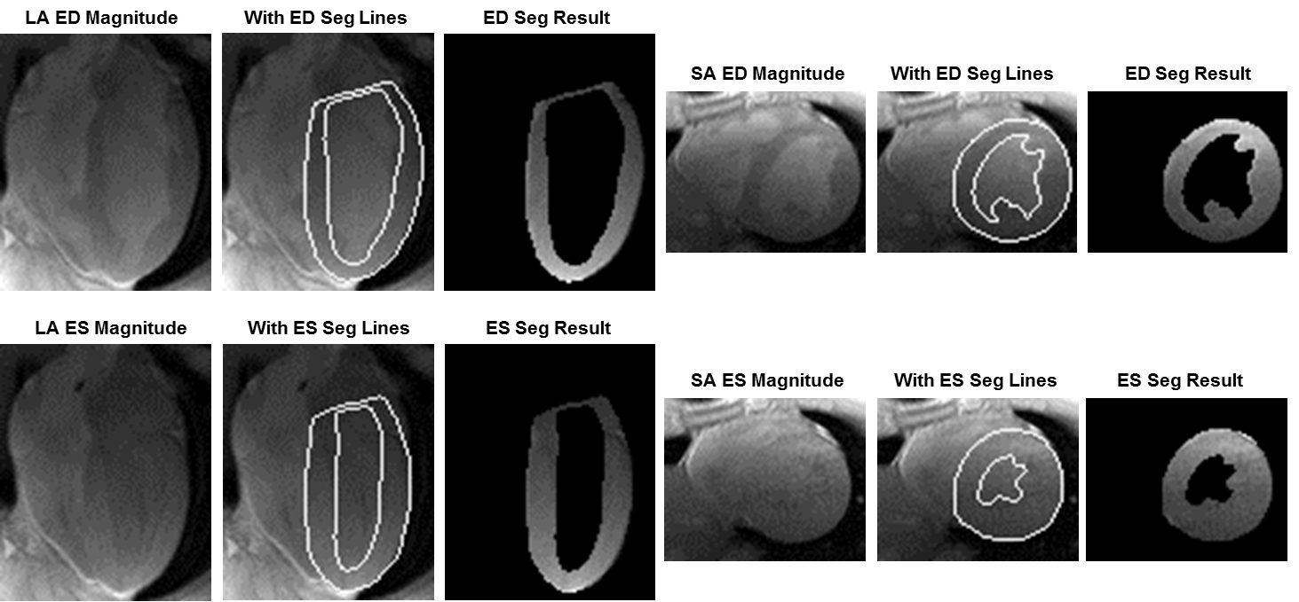
Figure 1 illustrates the ECG-triggered CWM sequence that acquires both normal and complimentary spatial modulation of magnetization (SPAMM/CSPAMM) prepared cine image sets. Before the SPAMM is enabled, a low-resolution pre-scan reference image is acquired for background phase correction. A classic “1-1” saturation SPAMM was modified were each non-selective rectangular RF pulse has a 45° flip angle. The cine readout uses a standard, low flip angle, spoiled GRE method with a parallel imaging acceleration factor of two. Within one breath-hold CWM acquires a full SPAMM image set then a full CSPAMM image set. CWM acquisitions were run with spatial modulation in the readout (RO) axis and phase encode (PE) axis directions to create orthogonal line-tags and combined for grid-tag patterns. Twelve healthy human subjects, 19-42 yo, 3 female, with informed consent, were scanned in a 3T Verio scanner (Siemens, Erlangen, DE) with a 32-chan anterior/posterior RF coil array (Invivo, Gainesville, FL). CWM scan slice locations included one long-axis (LA) 4-chamber and 1-3 mid-left ventricular (LV) short-axis (SA) views. Scan parameters included: field-of-view = 264x231 mm, slice thickness = 6 mm, matrix = 176x154, in-plane pixel size = 1.5x1.5 mm, flip angle = 8°, BW = 406 Hz/pix, averages = 1, cine phases = 20-25, and tag spacing = 6 mm. CWM image reconstruction was performed offline using custom Matlab programs (Mathworks, Natick, MA). Reconstruction included: 1) correct the SPAMM and CSPAMM background phase with the reference scan phase, 2) perform a complex image sum of the SPAMM+CSPAMM that eliminates the cosine modulation components to leave only the (central k-space) conventional T1-weighted component, from which, the magnitude images provide normal cine images without tags, 3) perform a complex image difference of SPAMM–CSPAMM that eliminates the central k-space T1-weighted component to leave only the cosine modulation components, from which, the phase images provide strong and persistent (0°/180°) phase-only tag lines that track cardiac displacement for strain calculation. Cardiac segmentation lines drawn onto CWM magnitude images also automatically segmented corresponding phase images (also Matlab). The cardiac strain analysis is described in detail in a separate submission. Briefly, cardiac strains were tracked by a novel non-linear least squares method combining quadratic b-spline contours, nearest neighbor optimized phase tracking, and physically-motivated regularizers. Cardiac LV SA, six sector, circumferential strain (Ecc) results were calculated and averaged around the LV.Results
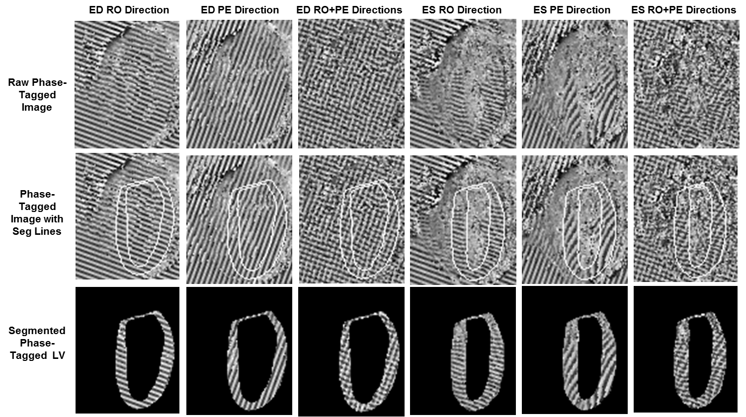
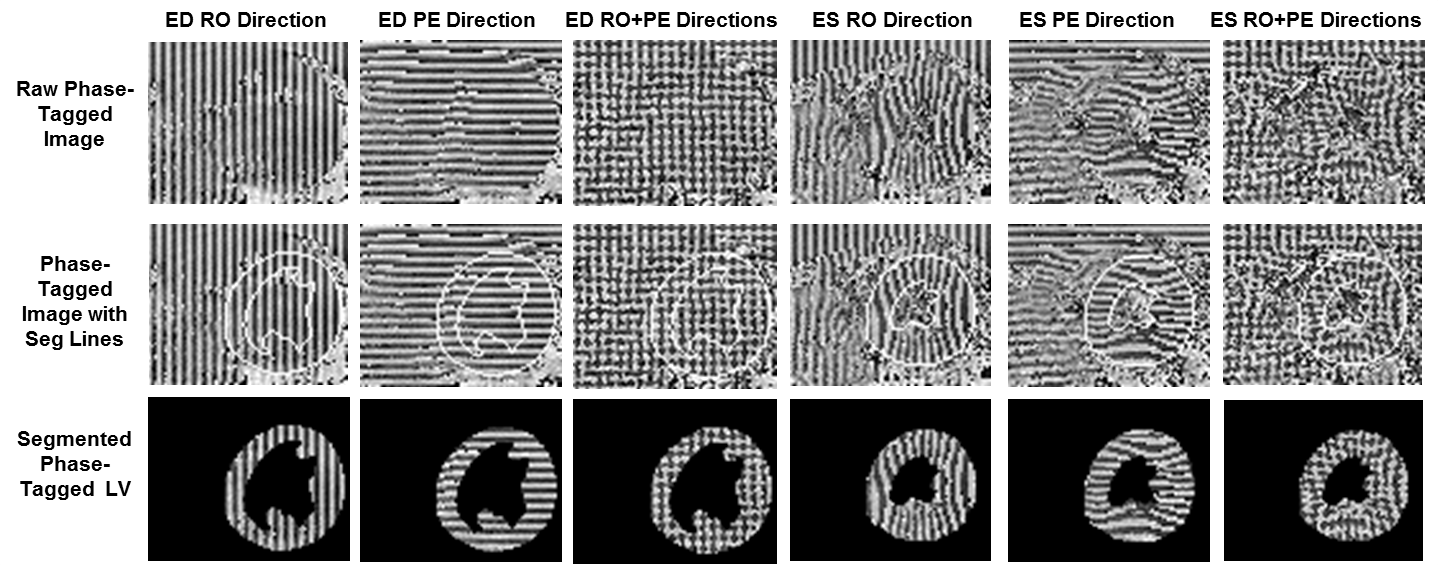
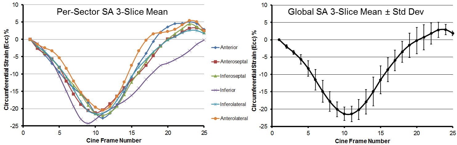
All CWM scans provided cine magnitude and spatial-modulated phase image sets suitable for strain analysis within a single breath-hold of 25 seconds. Figure 2 presents representative CWM LA and SA magnitude cine frames at end-diastole (ED) and end-systole (ES) time-points with subsequent LV segmentation. Figure 3 presents LA phase-tagged results at ED and ES with spatial-modulation applied in the RO, PE and combined RO+PE (grid) directions, where the segmented LV images show clear displacement and bending of the phase-tagging patterns at ES. Similar to Figure 3, Figure 4 presents SA phase-tagged results that also show clear displacement and bending of the LV phase-tagging patterns at ES. Figure 5 presents representative mean Ecc strain results averaged from 3 contiguous, mid-LV, SA slices for a human subject. All sectors, except the inferior sector, show nominal strain with consistent trajectories.Discussion
CWM can acquire cardiac cine and phase-tagged cine images in a single breath-hold. The integration of parallel imaging acceleration was essential to achieve one breath-hold per scan capability and the decoupled RO & PE encodings improved the strain analysis. The Ecc strain results generally agree with the previous literature5; however, further work is needed to improve tracking robustness and overall accuracy.Conclusions
Quantitative assessment of regional myocardial contraction is an essential element of diagnostic CMR. Cine “Watermark” MRI provides an approach to breath-hold cine imaging with integrated “hidden” capability to acquire strain data encoded into the phase. This work provides a platform for further sequence development to included further acceleration and the potential for SSFP imaging.Acknowledgements
Special appreciation to Martha Forloines for her valuable management of MRI operations and technical assistance.References
1. Bolster BD, et al. “Myocardial tagging in polar coordinates with striped tags”, Radiology 1990;177:769–72.
2. Axel L, Dougherty L. “MR imaging of motion with spatial modulation of magnetization”, Radiology 1989;171:841–9.
3. Axel L, Dougherty L. “Heart wall motion: Improved method of spatial modulation…”, Radiology 1989;172:349–50.
4. Edelstein WA, et al. “MRI: Time Is Dose - and Money and Versatility, J Am Coll Radiol 2010;7(8): 650–2.
5. Edvardsen T, et al. “Quantitative Assessment of Intrinsic Regional Myocardial
Deformation…”, Circulation 2002;106:50-56.
Figures