3141
Age-stratified normal left atrial deformation assessed by novel longitudinal strain parameters on a 3T MR scanner1National Heart Centre Singapore, Singapore, Singapore, 2Duke-NUS Medical School Singapore, Singapore, Singapore
Synopsis
The aim of this study was to examine the age-related normal left atrial (LA) longitudinal deformation by a novel and fast assessable strain parameter with standard cardiac magnetic resonance (CMR) imaging. A total of 60 heathy subjects (30 males, age between 20 and 80 years) were categorized by age into young, middle-aged, and elderly adults. The LA longitudinal strain and strain rate measurements corresponding to reservoir and conduit phases were negatively related to age, while no significant age-dependency was observed for LA contraction strain and strain rate values. These strain parameters can be used to evaluate LA deformation and functionality.
Background and Purpose
The left atrium (LA) is a significant determinant of cardiovascular morbidity and mortality. Its key role is to modulate left ventricular (LV) filling through reservoir, conduit and booster pump function1. Pressure-volume relationship remains the gold standard for assessment of LA function2, but this requires an invasive procedure. In clinical practice, echocardiography is the main diagnostic modality for LA structure and function through the use of volumetric analysis, tissue Doppler and atrial deformation imaging3. Cardiac magnetic resonance (CMR) and feature tracking offer the opportunity for non-invasive quantification of LA myocardial motion. Current study sought to investigate the age-stratified normal LA deformation assessed by novel strain measurements with cine CMR imaging.Methods
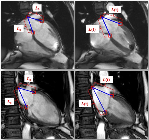
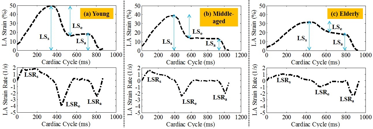
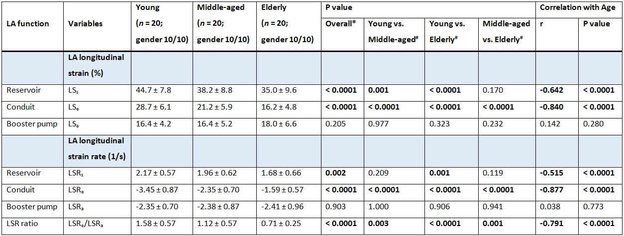
Sixty healthy volunteers without any signs, symptoms or history of cardiovascular diseases were examined in this study. Subjects were classified as “young” (range 20 – 35 years; gender male/female, 10/10 with age of mean ± SD, 26.8 ± 4.0/26.4 ± 3.3 years), “middle aged” (range 35 – 55 years; gender male/female, 10/10 with age of mean ± SD, 45.1 ± 5.8/44.4 ± 5.9 years), or “elderly” (range 55 – 80 years; gender male/female, 10/10 with age of mean ± SD, 68.3 ± 6.3/68.2 ± 6.9 years). CMR scan was performed on a 3T system (Ingenia, Philips Healthcare) using balanced turbo field echo sequence. End-expiratory breath hold cine images were acquired in multi-planar short- and long-axis views. An in-house developed program4,5 was applied to semi-automatically track the atrioventricular junction (AVJ) points and the LA apex in four- and two-chamber views. The long-axis LA longitudinal strain was calculated as $$$LS(t)=\frac{(L(t)-L_{0})}{L_{0}}$$$, with $$$LS(t)$$$ the longitudinal strain, $$$L_{0}$$$ the initial length, and $$$L(t)$$$ the length at cardiac time $$$t$$$ (Fig. 1). The reservoir strain LSs, conduit strain LSe and contraction (booster pump) strain LSa were identified from the strain curve (Fig. 2 top); the longitudinal strain rates LSRs, LSRe and LSRa during the three LA phases were obtained from the strain rate curve (Fig. 2 bottom). Data was analyzed using SPSS (version 17.0, Chicago, IL, USA). An F-test was used to test the omnibus hypothesis of equality among age groups, which was then followed up with post hoc Tukey or Games Howell test between each group pair. A P value < 0.05 was considered statistically significant.Results
Analysis was feasible for all subjects with < 5 min per case. Figure 2 showed the global LA strain and strain rate curves by averaging all strain values obtained in two- and four-chamber views for a 26-yr-old young female, 35-yr-old middle-aged female, and 78-yr-old elderly female healthy controls. The age-stratified strain and strain rate values were shown in Table 1. The global reservoir and conduit strains were inversely and significantly correlated with age (LSs: r = -0.642, LSe: r = -0.840, both P < 0.0001). Accordingly, the difference between the young and elderly group yielded significance for LSs and LSe. The global strain rates during reservoir (LSRs) and conduit (LSRe) phases, as well as the strain rate ratio LSRe/LSRa decreased significantly with age (r = -0.515, -0.877, and -0.791, respectively, all P < 0.0001). The group of young subjects had significant higher global LSRe and ratio value LSRe/LSRa in comparison with middle-aged and elderly groups. The strain and strain rate values during contraction phase were not associated with age. No significant differences for LSa and LSRa were observed among the age groups. The intra- and inter-observer reproducibility of current CMR-based method was excellent for all strain and strain rate measurements with small limits of disagreement and coefficient of variation (< 5%).Conclusions
Current study presented fast assessable and highly reproducible LA longitudinal strain and strain rate measurements stratified by age. The LA deformation during reservoir and conduit phases had inverse relationship with respect to age in healthy subjects.Acknowledgements
No acknowledgement found.References
1. Hoit BD. Left atrial size and function: role in prognosis. J Am Coll Cardiol. 2014;63:493-505.
2. Rosca M, Lancellotti P, Popescu BA, Piérard LA. Left atrial function: pathophysiology, echocardiographic assessment, and clinical applications. Heart. 2011;97(23):1982-1989.
3. Blume GG, Mcleod CJ, Barnes ME, et al. Left atrial function: physiology, assessment, and clinical implications. Eur J Echocardiogr. 2011;12(6):421-430.
4. Leng S, Zhao XD, Huang FQ, et al. Automated quantitative assessment of cardiovascular magnetic resonance-derived atrioventricular junction velocities. Am J Physiol Heart Circ Physiol. 2015;309:H1923-H1935.
5. Leng S, Jiang M, Zhao XD, et al. Three-dimensional tricuspid annular motion analysis from cardiac magnetic resonance feature-tracking. Ann Biomed Eng. 2016 [Epub ahead of print].
Figures