3140
Using Non-Contrast-Enhanced Magnetic Resonance Angiography (MRA) in Evaluating Preoperative Facial Artery of Vacularized Submental Lymph Node Flap: Compared to Conventional Contrast-Enhanced MRA1Department of Medical Imaging and Intervention, Chang Gung Memorial Hospital, Taoyuan, Taiwan, 2GE Healthcare, Taiwan, 3GE Healthcare MR Research China, Beijing, People's Republic of China
Synopsis
The aim of this study was to determine whether the recently
developed non-contrast-enhanced Inhance 3D velocity magnetic resonance
angiography (Inhance 3D MRA) technique is capable of evaluating the course of facial
artery (branch artery) in preoperative evaluation of vacularized submental
lymph node flap. The result shows that Inhance 3D MRA is a promising method for
assessing facial artery course without concerning the contrast media (CM) travel time and the venous contamination.
Purpose
The course and morphology of facial artery are important for preoperation evaluation of submental vascularized lymph node flap transfer for patients with lymphedema1. Facial artery is a main branch of external carotid artery and it is seldom evaluated by MRA. CE-MRA has been proven to be useful for visualizing major arteries in the head and neck such as carotid artery and basilar artery in clinical setting2. However, it is difficult to apply the same measurement and to find the optimal acquistion timing in assessing facial artery due to variable travel time of contrast media (CM) in the neck. Furthermore, recent reports3 linking the use of gadolinium-based MR CM with nephrogenic systemic fibrosis have hampered their widespread application in patients with renal insufficiency. A novel non-contrast MRA technique using phase contrast-based (PC) 3D velocity MRA4 (Inhance 3D MRA) was recently proposed. Unlike traditional PC-MRA, Inhance 3D MRA is compatible with parallel imaging and utilizes time-efficient elliptical k-space filling technique. The purpose of this study is to determine the performance of Inhance 3D MRA in the depiction of the facial arteries for preoperative planning and compared the result with that in CE-MRA.Method and Material
Twenty patients with lymphedema were imaged in a 3T clinical
scanner (Discovery MR750, GE Healthcare, Milwaukee, USA ) using an 8-channel
brain coil as the signal detection and whole body coil for RF transmission.
Images from Inhance 3D
MRA and CE-MRA were acquired with the matched slice coverage and
resolution (FOV=300 mm; matrix=352×224) and other scanning parameters were as
follows: TR/TE= 8.9/3.9 msec and 3.9/1.4 msec for Inhance 3D MRA and CE-MRA,
respectively. For better depicting the course of facial artery, the facial
anatomical reference and neighboring position of lymph node were acquired by
sagittal Cube T1-weighted images (T1WI) (TR/TE= 600/13 msec) and fat-saturated
Cube T2-weighted images (T2WI) (TR/TE= 2500/76 msec), respectively. The 18
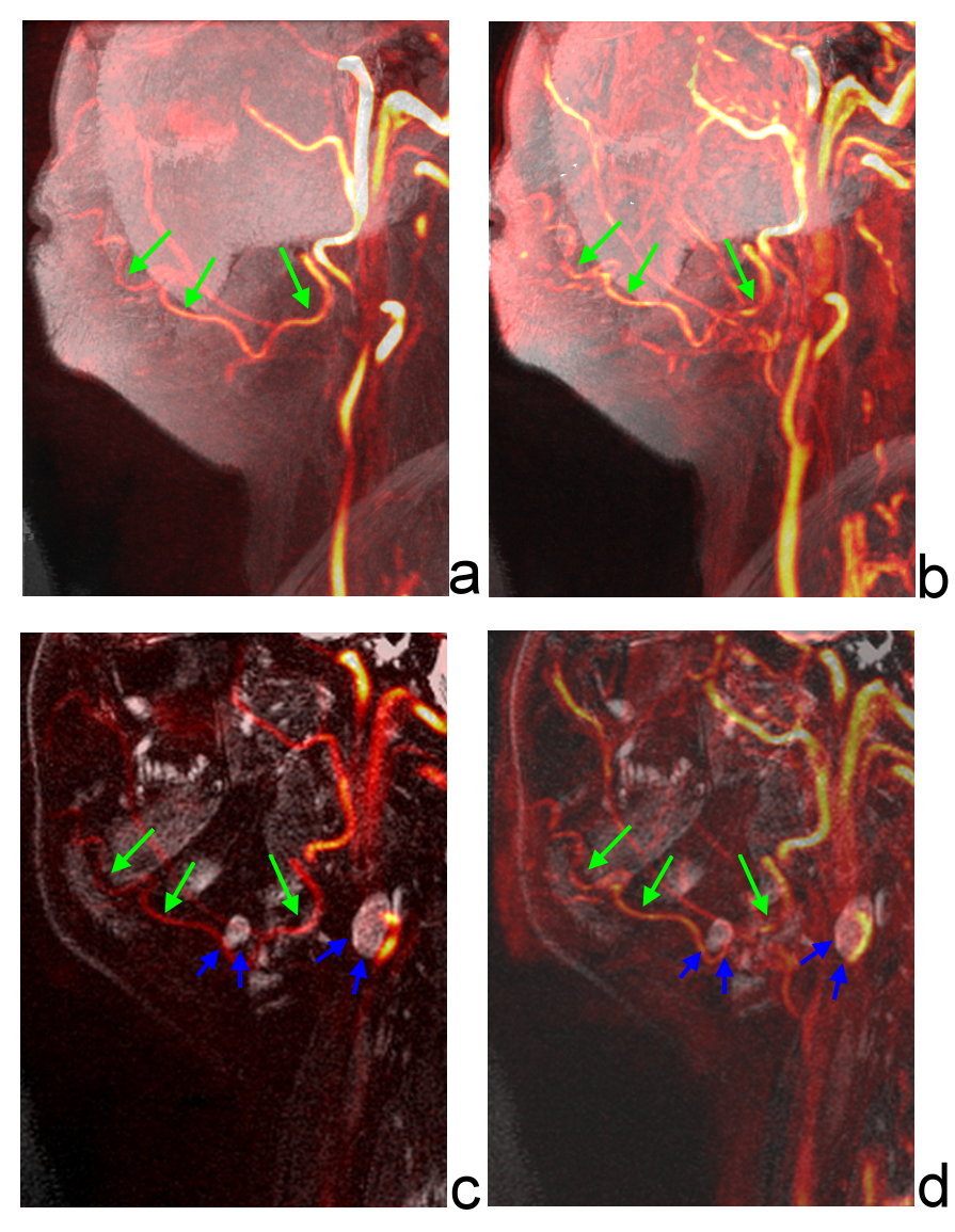
mm-thick slabs were generated by maximum intensity projection (MIP) of Inhance
3D MRA or CE-MRA fused with Cube T1WI or fat-saturated Cube T2WI (Figure 1). The
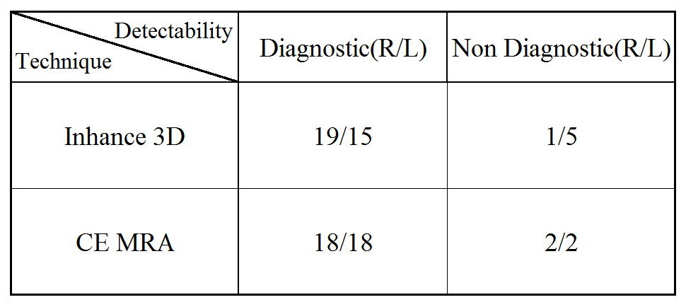
visualibilty of facial artery at two MRA techniques was assessed by two radiologists and rated
on four-point score: poor (0), questionable (1), adequate (2) and good (3). To
determine the diagnostic ability for clinical application, the results were
dichotomized into diagnostic (score 3 and 2) and non-diagnostic (score 1 and
0). The significance differences between the diagnostic and non-diagnostic in MRA
results generated by Inhance 3D MRA and CE-MRA were evaluated with the Fisher
Exact test.Result and Discussion
Depending on the results of image score dichotomized
into
diagnostic and non-diagnostic in two MRA techniques (Table 1), there was no
significant difference in the diagnostic ability between Inhance 3D MRA and
CE-MRA (p =0.447 and 1 for left and right side facial artery,
respectively). A representative morphology of left facial artery was
visualized in both Inhance 3D MRA (Figure 1a) and CE-MRA (Figure 1b). Fused MRA
with fat-saturated 3D Cube T2WI (Figure 1c and 1d), the lymph node position can
be located. This would be helpful for platic surgeon to do preoperative
planning. The acquisition timing for CE-MRA has been optimized by finding
maximal of CM travel time in evaluating facial artery. Apparently, CE-MRA
showed high vessel signal due to the presence of CM compared to that in Inhance
3D MRA. However, the enhanced signal in background tissue caused by venous
contamination was also found in CE-MRA (Figure 1b). Unlike CE-MRA, Inhance 3D MRA
provides the excellent contrast between background tissue and facial artery.Conclusion
Our study demonstrated that Inhance 3D MRA is a reliable method for evaluating preoperative facial artery course without concerning the travel time of CM and it could be an alternative to CE-MRA in patient with renal insufficiency. Non-contrast Inhance 3D MRA is expected to have wide clinical application and diagnostic value in the visualization of small artery.Acknowledgements
No acknowledgement found.References
1. Cheng MH, Huang JJ, Nguyen DH, et al. A noval approach to the treatment of lower extremity lymphedema by transferring a vascularized submental lymph node flap to the ankle. Gynecol Oncol. 2012;126(1):93-8
2. Leclerc X, Gauvrit JY, Nicol L, et al. Contrast-enhanced MR angiography of the craniocervical vessels : a review. Neuroradiology 1999;41:867–874.
3. Broome Dale R. Nephrogenic systemic fibrosis associated with gadolinium based contrast agents: A summary of the medical literature reporting. European Journal of Radiology 2008;66:230–234.
4. Nina L, Tobias BB, Juergen L, et al. Evaluation of the supraaortic arteries using non-contrast-enhanced Velocity MR Angiography “Inhance”. Neuroradiology 2012;54:1215–1219.
Figures