3134
Flow enhanced (FEED) Arterial Spin Labeling Angiography with segmented Zero Echo Time Readout1MR Research China, GE Healthcare, Beijing, People's Republic of China
Synopsis
ASL based MRA with continuous labeling has the optimal label efficiency. To achieve data acquisition efficiency, a long readout train is frequently used after labeling. Flow void might appear due to the lack of tagged blood entering the imaging volume during readout. A method named hybrid ASL address this problem with additional pulsed inversion. Yet, blood inverted by pulsed ASL still experiences considerable T1 decay during readout. We propose a method termed flow enhanced ASL (FEED). Interleaved labeling modules along with segmented readout was employed. As the results show, FEED could increase the signal level without prolonging scan time.
Purpose
Magnetic resonance imaging is being widely used for imaging the cerebral vasculature as an alternative to digital subtraction angiography (DSA) or computational tomography angiography (CTA). Time of flight (TOF) imaging is the most common method, but has several limitations including vulnerability to susceptibility sources, hemodynamic imperfections, and limited spatial coverage. Contrast enhanced MRA may overcome the shortcomings of TOF but requires contrast administration. Arterial spin labeling (ASL) angiography uses RF labeled arterial blood as endogenous contrast and has been demonstrated to feature the advantages of CE MRA while being contrast free. There are three common types of labeling strategies: pulsed ASL(pASL), pseudo-continuous ASL(pcASL), continuous ASL(cASL). Continuous ASL features the highest labeling efficiency, and the following zero TE acquisition method minimizes the T2 decay and conserves high level of signal fidelity. However, to achieve data acquisition efficiency in practice, a relatively long readout train is needed after labeling, which lead to considerable magnetization T1 decay and may result in flow void in the resulting angiogram [1]. A past technique named hybrid ASL (hASL) [2] uses an additional pulsed inversion for signal loss compensation. In this work, we propose and investigate a new method named flow enhanced (FEED) ASL to further improve for blood signal in the angiogram.Method
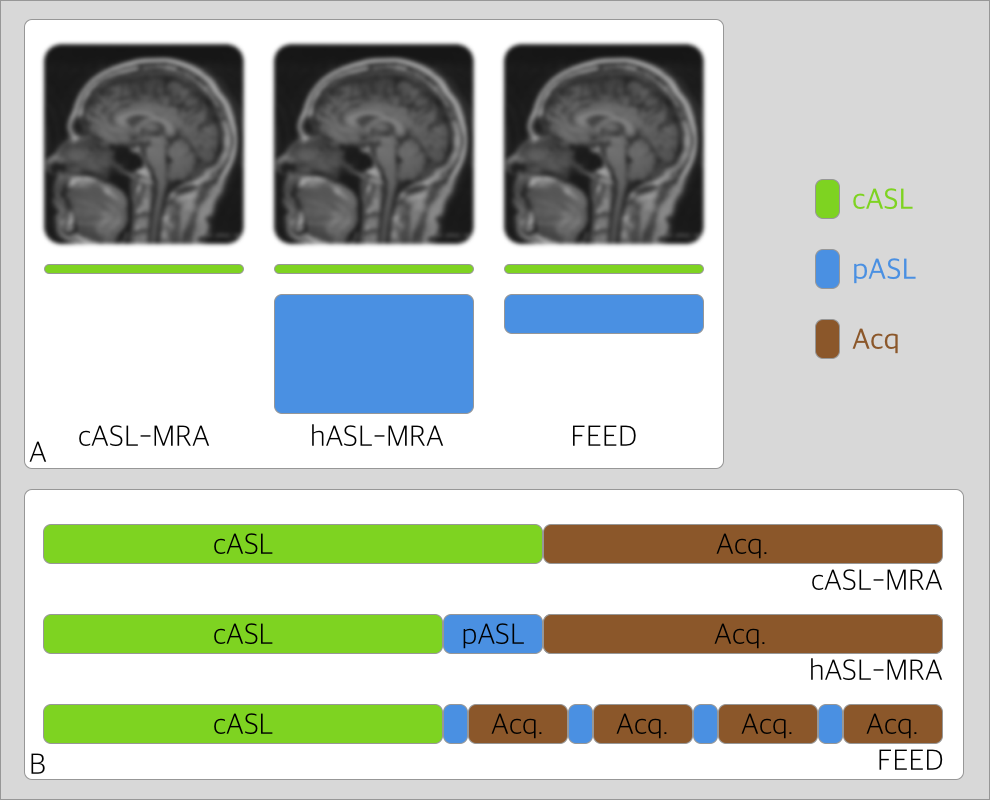
A comparison of the pulse sequences of cASL, hASL, and the proposed FEED ASL is demonstrated in Fig. 1. Continuous ASL labels a thin slab beneath the imaging volume, and a readout train immediately follows the labeling module. However, fresh uninverted or partially inverted blood may flow into the imaging volume during the readout leading to flow void in the early section of the angiogram. Hybrid ASL adds a thick slab inversion after the continuous labeling so that blood inverted by the pulsed inversion will be present during the readout train to compensate for the potential signal loss. FEED ASL adds interleaved labeling modules along with segmented readout train with a moderate slab inversion. In this way, the effects of magnetization T1 decay during the readout may be further reduced. To test the performance of the proposed method, cASL, hASL and FEED ASL were implemented with the same total labeling duration of 1.4s: the labeling duration was 1.4s in cASL; the continuous labeling duration was 1.2s and pulsed labeling was 200ms in hASL; the continuous labeling duration was 1.2s and 4 pulsed labeling modules of 50ms each was implemented in FEED ASL. The inversion slab thickness was proportionally reduced in FEED ASL that it was 1/4 of that in hASL, which is to avoid the case of double inverting of blood so that no effective inversion was made. The same zero TE readout train of 512 spokes was used in cASL and hASL, whereas FEED ASL used 4 readout train segments with 128 spokes each. The other pulse sequence parameters are as following: flip angle of 3 degrees, FOV of 160 mm, and 1mm isotropic resolution. A healthy volunteer was recruited for this study with consent form obtained. The resulting images were compared both qualitatively and quantitatively.Results
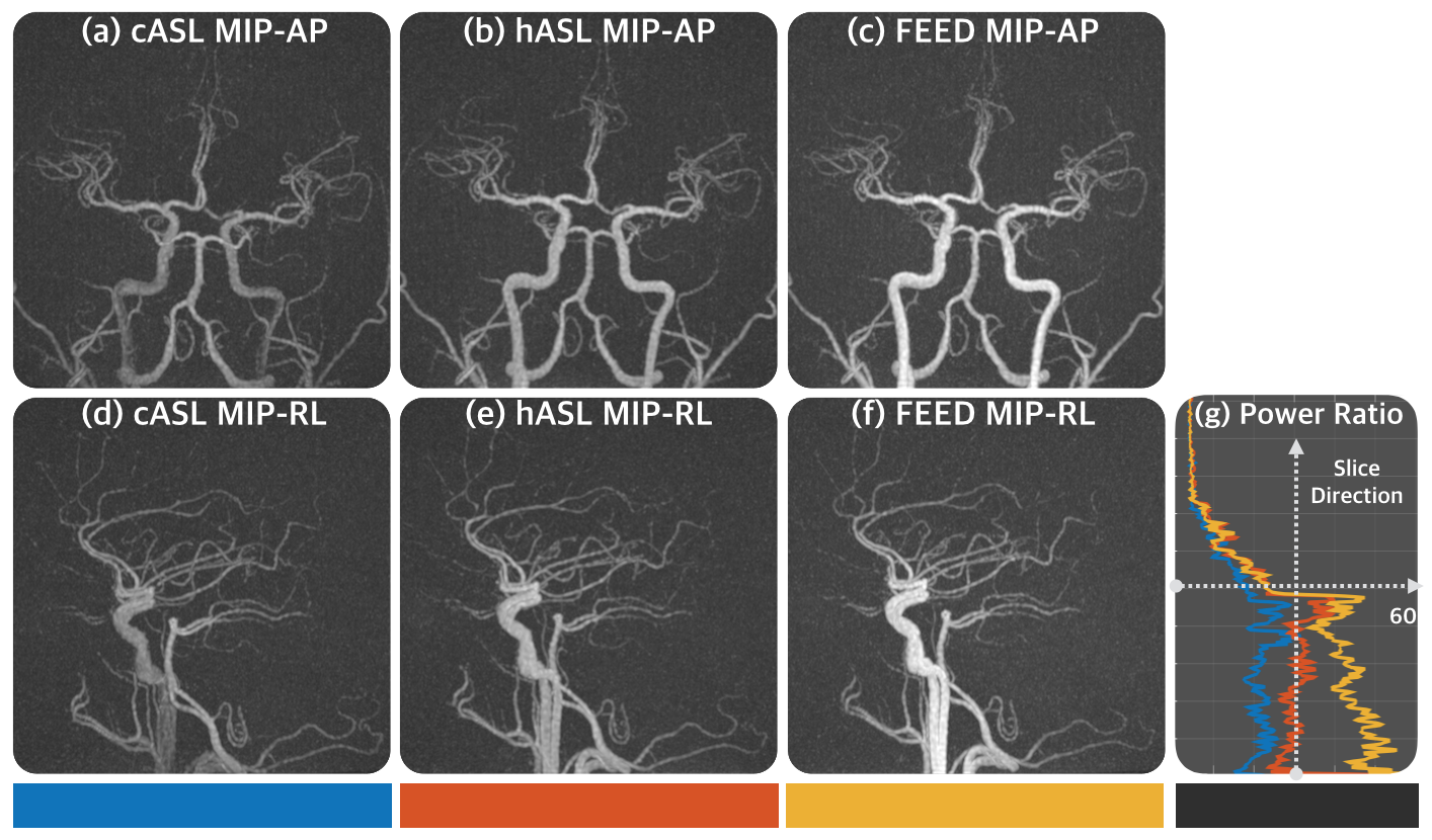
The resulting maximum intensity projection images of cASL, hASL, and FEED ASL are shown in Fig.2 (with the same window leveling). It can be seen that the flow void in cASL is obvious that the entry terminal of carotid artery is nearly hollow (Fig.2 a,d), whereas the flow void is effectively compensated by the pulsed inversion in hASL (Fig.2 b,e). FEED ASL further improves the integrity of the angiogram that more homogenous vascular structures are obtained (Fig.2 c,f). Fig.2.g shows the ratio of level of maximum signal power over the level of noise power along the slice direction (inferior to superior), it can be seen that an apparently higher level of signal was received in FEED ASL at the inferior end as compared to that of cASL and hASL.Discussion and Conclusion
In this work, a new ASL MRA method that employs interleaved labeling model was proposed on the basis of previously proposed hASL. It effectively improves blood signal level and the integrity of the vascular structure as compared to previous methods. This method does not require extra labeling time nor prolongs the readout train length for the added benefits, as demonstrated in this preliminary investigation. A further area for optimization is the choice of inversion slab thickness in FEED method, as a slab to thin may still lead to uninverted blood whereas a slab too thick may result in twice inversion that would again become uninverted.Acknowledgements
No acknowledgement found.References
[1] I.Koktzoglou, etc. ASL Carotid MRA: A Phantom Study Examing the Impact of Technical an d Hemodynamic Factors, MRM, 2015.
[2] I.Koktzoglou, etc. Nonenhanced hybridized ASL MRA of the extraranil carotid arteries using a fast low angle shot readout at 3T, JCMR, 2016
Figures