3131
Super-Resolution Intracranial Quiescent-Interval Slice-Selective Magnetic Resonance Angiography1Radiology, NorthShore University HealthSystem, Evanston, IL, United States, 2Radiology, University of Chicago Pritzker School of Medicine, Chicago, IL, United States, 3Siemens Healthineers, Chicago, IL, United States, 4Radiology, Northwestern University Feinberg School of Medicine, Chicago, IL, United States
Synopsis
We evaluated whether super-resolution reconstruction could be used to improve the vascular detail of quiescent-interval slice-selective (QISS) MRA of the intracranial circulation. Results suggest that super-resolution reconstruction can substantially improve intracranial arterial delineation without the signal-to-noise ratio penalty of directly acquiring thin-slice images.
Introduction
Quiescent-interval slice-selective (QISS) magnetic
resonance angiography (MRA) is a multi-slice 2D technique that has been
reported for the evaluation of the peripheral, renal, carotid and coronary
arteries.1-3 However, a potential drawback of 2D MRI methods such as
QISS is limited spatial resolution in the slice-select direction due to
practical concerns of signal-to-noise ratio, gradient slew rates and signal
from out-of-slice vascular spins. Depending on the application, the typical
slice thickness used with QISS MRA ranges from 1.5mm to 3 mm. Well-established
in the signal processing literature, “super-resolution” is an approach for creating
a higher-resolution image from a series of overlapping and offset lower-resolution
images.4,5 We hypothesized that super-resolution reconstruction could
be used to improve the slice resolution of QISS MRA, and sought to apply this
strategy for imaging the intracranial circulation.Methods
This prospective study was approved by our institutional review board. A prototype implementation of QISS MRA was used to image the intracranial arteries of healthy subjects (n=14, age range=24-53) on 1.5T and/or 3T MR systems (MAGNETOM Avanto, Verio and Skyrafit, Siemens Healthcare). Typical imaging parameters for the prototype QISS sequence included: a flow-compensated fast low-angle shot (FLASH) readout, TR/TE/flip = 10ms/5ms/30°, 0.7mm×0.7mm-1.0mm×1.0mm in-plane spatial resolution interpolated to 0.35mm×0.35mm-0.5mm×0.5mm, 1.5mm slices acquired with 50%-67% overlap, inflow times of ~350ms, 120-128 slices, 1-3 shots per slice, scan times of ~1.5min-4.5min. Super-resolution reconstruction was implemented in the manner of Irani-Peleg.6 Quantitative analyses were performed in the middle cerebral artery; analyses included the calculation of arterial sharpness, arterial full-width-at-half-maximum, and arterial-to-background contrast-to-noise ratio. Preliminary comparisons were made with 3D time-of-flight MRA matched for spatial resolution and scan time.Results
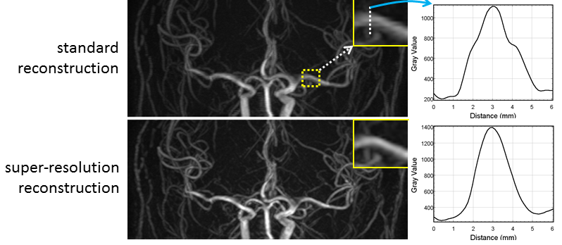
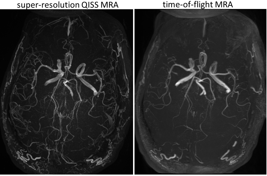
Compared with standard reconstruction, super-resolution reconstruction resulted in visible improvement in arterial delineation in the slice-select direction (Figure 1), a 17% improvement in arterial sharpness (P<0.001), and an 11% reduction of arterial full-width-at-half-maximum (P<0.001). Contrast-to-noise ratio in the reconstructed super-resolution images was significantly improved at 3T versus 1.5T (P<0.001), and was not degraded with respect to values obtained with standard reconstruction. Intracranial QISS MRA routinely matched or surpassed 3D time-of-flight MRA in intracranial arterial detail, and demonstrated an improved degree of background signal suppression (Figure 2). Single-shot QISS MRA could provide 9.6cm of axial coverage in as little as 90 seconds.Discussion and Conclusion
Super-resolution reconstruction is a useful method
for improving the reconstructed slice resolution when applied to QISS MRA of
the intracranial circulation. Quantitative analyses suggest that
super-resolution reconstruction can improve arterial delineation without the signal-to-noise
ratio penalty of directly acquiring thin-slice images. Although the present
study focused on QISS MRA of the intracranial circulation, we anticipate that
super-resolution reconstruction can be applied to QISS MRA of carotid, renal, coronary
and great vessels.Acknowledgements
Study was funded in part by NIH grants R01 HL130093 and R21 HL126015.References
1. Edelman RR, Sheehan JJ, Dunkle E, Schindler N, Carr J, Koktzoglou I. Quiescent-interval single-shot unenhanced magnetic resonance angiography of peripheral vascular disease: Technical considerations and clinical feasibility. Magn Reson Med. 2010 Apr;63(4):951-8.
2. Koktzoglou I, Murphy IG, Giri S, Edelman RR. Quiescent interval low angle shot magnetic resonance angiography of the extracranial carotid arteries. Magn Reson Med. 2016 May;75(5):2072-7.
3. Edelman RR, Giri S, Pursnani A, Botelho MP, Li W, Koktzoglou I. Breath-hold imaging of the coronary arteries using Quiescent-Interval Slice-Selective (QISS) magnetic resonance angiography: pilot study at 1.5 Tesla and 3 Tesla. J Cardiovasc Magn Reson. 2015 Nov 23;17:101.
4. Park SC, Park MK, Kang MG. Super-resolution image reconstruction: a technical overview. IEEE signal processing magazine. 2003 May;20(3):21-36.
5. Greenspan H, Oz G, Kiryati N, Peled S. MRI inter-slice reconstruction using super-resolution. Magnetic resonance imaging. 2002 Jun 30;20(5):437-46.
6. Irani M, Peleg S. Improving resolution by image registration. CVGIP: Graphical models and image processing. 1991 May 31;53(3):231-9.
Figures