3035
Transport of Hyperpolarized Nanodiamonds1ARC Centre of Excellence for Engineered Quantum Systems, School of Physics, University of Sydney, Sydney, Australia
Synopsis
Hyperpolarized 13C MRI using nanodiamond (ND) holds the potential for tailored diagnostic imaging combined with targeted drug delivery in the human body. An obstacle to realizing this potential is the transfer of hyperpolarized ND from the hyperpolarizer to the patient without losing the majority of the 13C polarization as it travels through low magnetic fields before reaching the MRI scanner. We demonstrate that polarization loss is highly dependent on magnetic field and construct a system of transfer magnets that improves the transfer efficiency of our hyperpolarized ND by more than an order of magnitude.
Purpose
Nanodiamond (ND) is a biocompatible material1 that has shown promise as a drug delivery platform2,3 and an optically-trackable bioprobe inside living cells.4 It has also been shown that ND can be hyperpolarized5,6 and exhibits long spin-lattice relaxation times up to the order of one hour.7 Thus, ND has the potential to take advantage of the increase of signal to noise of four orders of magnitude provided by carrying out dynamic nuclear polarization (DNP) at low temperature and high field before shuttling samples to room temperature for acquisition.8 However, these advantages of ND are of no use in the context of clinical MRI if the hyperpolarization does not survive the transition from the hyperpolarizer into a patient inside an MRI machine. We have found the polarization of our ND quenches at significantly higher fields than those often used successfully to transfer solutions in dissolution DNP experiments. Those working with dissolution DNP have sought to optimize transfer efficiency by placing permanent magnets around the tube carrying the hyperpolarized fluid.9 Here we extend that approach to the more strict demands of ND.Methods
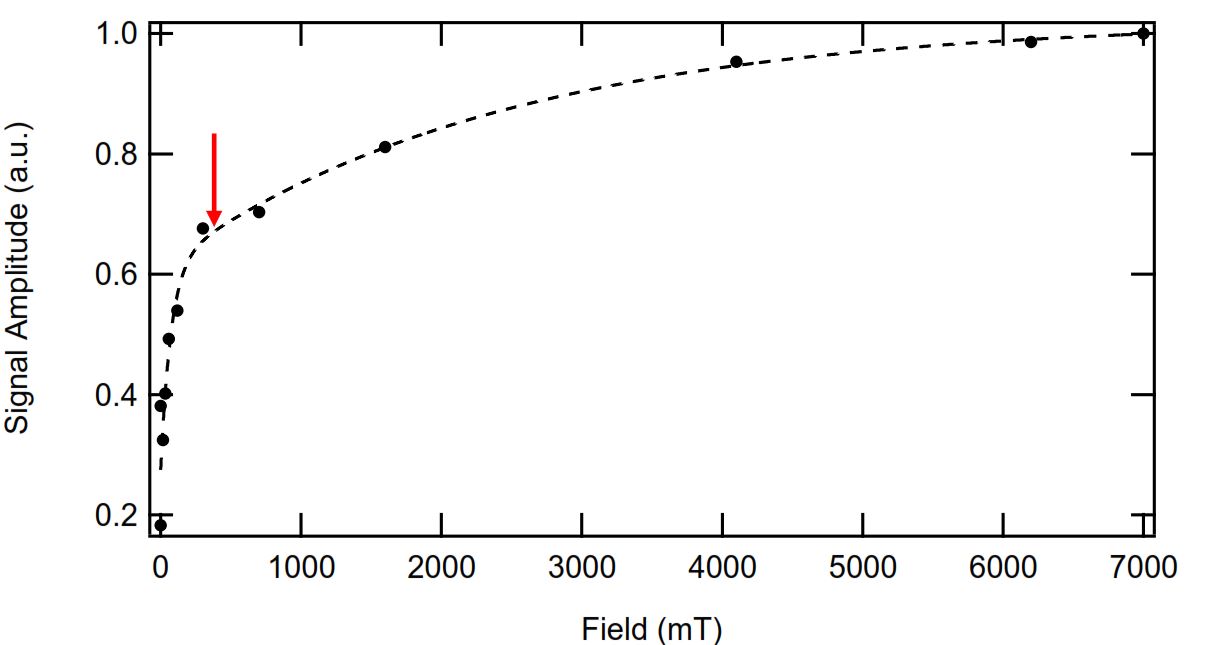
Experiments to gauge the loss of 13C polarization during a short transfer as a function of magnetic field were carried out with a 7 T NMR magnet at room temperature. For each run a 2 μm median particle size ND powder (Microdiamant MSY 1.5-2.5) was allowed to build up thermal polarization for 20 minutes before being lifted to a position of lower magnetic field for 20 seconds then lowered back into the NMR coil at 7 T for immediate signal acquisition with a CPMG pulse train. For a near-zero magnetic field the sample was lifted out of the NMR magnet and placed in a μ-metal chamber.
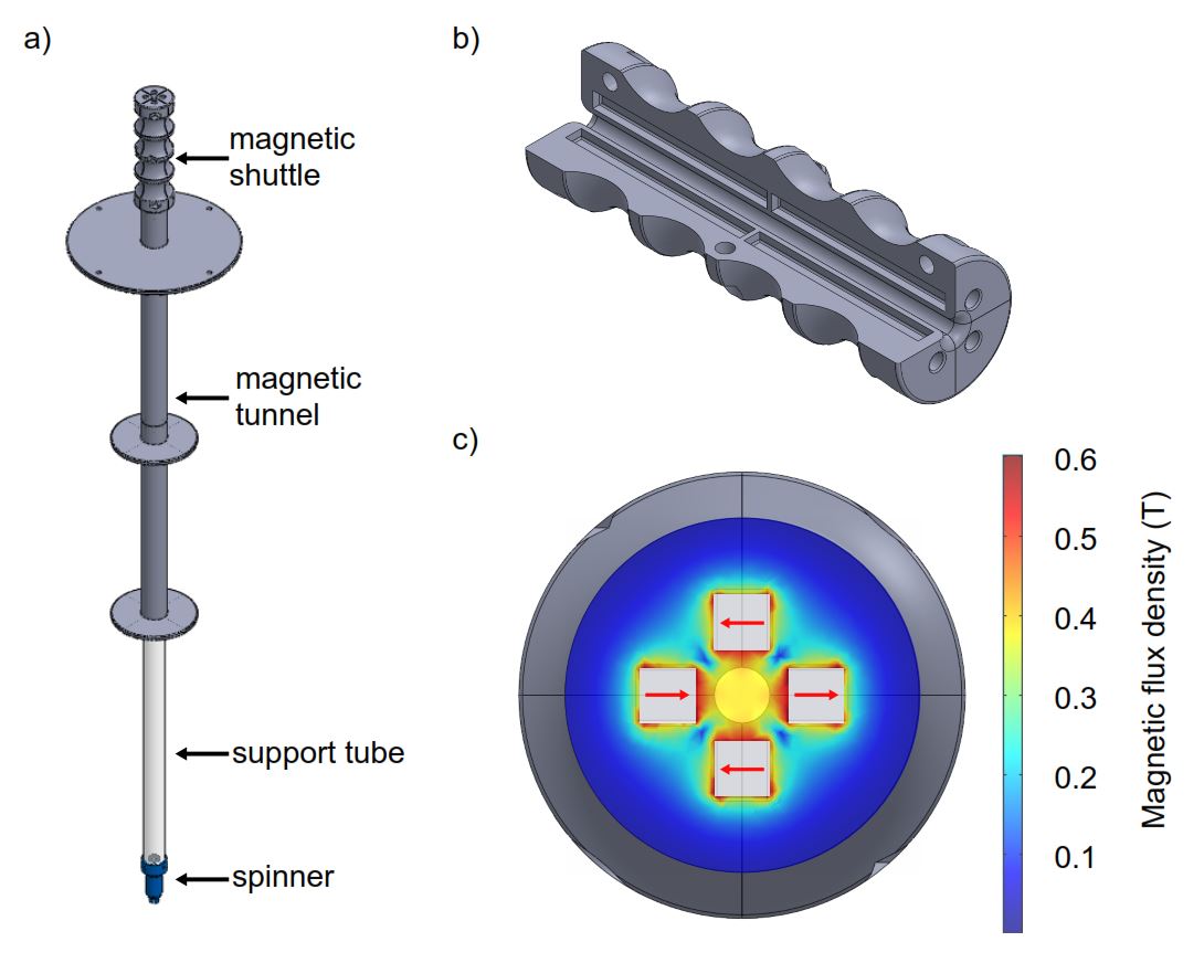
For the hyperpolarized transfer experiments a 2 μm ND sample was hyperpolarized with 80.895 GHz microwaves in a home-built DNP probe at 2.88 T, 6.5 K for 20 minutes before being transferred to a shielded 7 T micro-imager for signal acquisition with a single π/2 pulse. A series of Halbach arrays10 were positioned around the top of the cryostat containing the DNP probe, a magnetic tunnel consisting of a simplified Halbach array was placed in the shielded 7 T micro-imager and a transfer magnet was used to shuttle the sample between the polarizer and imager (see Figure 1). The magnetic field experienced by the sample never drops below 10 mT and is above 100 mT for most of the transfer time of < 10 seconds.
Results
Depolarization results are shown in Figure 2. The signal remaining decreases with magnetic field and shows behaviour characterised by two distinct regions. For fields above 300 mT the majority of the signal remains after 20 seconds. Below 300 mT depolarization rates increase dramatically.
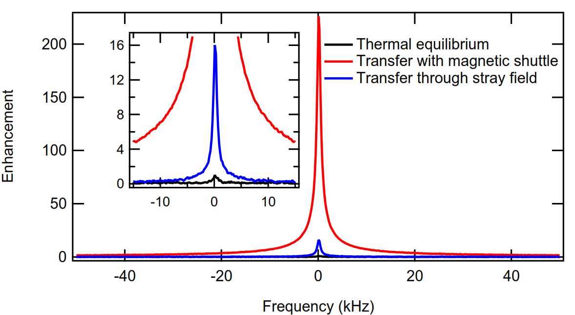
Transfer experiment results (see Figure 3) show the clear advantage of transfer with permanent magnets limiting the time the sample spends at low field. The transfer carried out with the transfer magnet is more than 14 times better than the transfer carried out where the sample travelled through the stray magnetic fields between the polarizer and imaging magnet. The enhancement of 226 for the transfer with the magnetic shuttle corresponds to a 13C polarization of 0.135% in the micro-imager. The polarization of the same sample in the polarizer before transfer was 3.2%.
Discussion
From the depolarization experiment we infer that a field of a few Tesla is necessary to transport hyperpolarized ND with minimum signal loss, but to maintain the majority of the polarization a field of greater than 300 mT is sufficient.
The hyperpolarized transfer experiments support
our inferences regarding the fields required to more efficiently transport our
hyperpolarized ND samples. However, the enhancement numbers indicate
significant room for improvement as they show that as little as 4% of the
polarization achieved in the polarizer survives after transfer to the imager. We are currently constructing a new DNP probe incorporating
a magnetic tunnel to keep the field experienced by our ND > 400 mT. We anticipate this will gain us another order of magnitude in
transfer efficiency.
Conclusion
The depolarization and transfer experiments presented here suggest that although limiting the polarization loss during transport of hyperpolarized ND samples is an obstacle to the application of ND for hyperpolarized 13C MRI it is by no means insurmountable. There are a number of paths open for future work to improve transfer efficiency, including incorporating a magnetic tunnel within future DNP probe designs, selecting ND with more favorable T1 field dependence characteristics and using a magnet with two isocenters.11Acknowledgements
This work was supported by the Australian Research Council Centre of Excellence Scheme (Grant No. EQus CE110001013) ARC DP1094439 and the Lockheed Martin Corporation.References
1. Zhu Y, et al. The Biocompatibility of Nanodiamonds and Their Application in Drug Delivery Systems. Theranostics 2012;2(3):302-312.
2. Li J, et al. Nanodiamonds as intracellular transporters of chemotherapeutic drug. Biomaterials. 2010 Nov;31(32):8410-8.
3. Chow EK, et al. Nanodiamond therapeutic delivery agents mediate enhanced chemoresistant tumor treatment. Sci Transl Med. 2011 Mar 9;3(73):73ra21.
4. McGuinness L. P, et al. Quantum measurement and orientation tracking of fluorescent nanodiamonds inside living cells. Nat Nanotechnol. 2011 May 8;6(6):358-63.
5. Dutta P, et al. Nanodiamond as a New Hyperpolarizing Agent and Its (13)C MRS. J Phys Chem Lett. 2014 Feb 6;5(3):597-600.
6. Casabianca L. B, et al. Factors Affecting DNP NMR in Polycrystalline Diamond Samples, J. Phys Chem C 2011 115 (39), 19041-19048.
7. Rej E., et al. Hyperpolarized nanodiamond with long spin-relaxation times. Nat Comms, 2015 6, 8459.
8. Ardenkjær-Larsen J. H, et al. Increase in signal-to-noise ratio of >10,000 times in liquid-state NMR. PNAS 2003 October;100(18):10158-63.
9. Milani J, et al. A magnetic tunnel to shelter hyperpolarized fluids, Rev. Sci. Instrum. 2015;86, 024101.
10. Halbach K, Design of permanent multipole magnets with oriented rare earth cobalt material, Nuclear Instruments and Methods, 1980;169(1):1-10.
11. Leggett J, A dedicated spectrometer for dissolution DNP NMR spectroscopy. Phys. Chem. Chem. Phys. 2010;12:5883-5892.
Figures