3010
Improved Triple-refocusing 1H MRS at 3T for detection of GABA and Glutamate in human brain in vivo1Advanced Imaging Research Center, University of Texas Southwestern Medical Center, Dallas, TX, United States
Synopsis
Reliable detection of GABA is important for studies in neuro-psychiatric diseases. In vivo 1H GABA resonances are extensively overlapped with the neighboring resonances including glutamate and glutamine. We present a new triple-focusing 1H MRS method which can fully resolve GABA 2.29-ppm and Glu 2.35 ppm signals at 3T.
PURPOSE
Alternations in the concentrations of primary inhibitory neurotransmitter, γ-aminobutyric acid (GABA), and major excitatory neurotransmitter, Glutamate (Glu), in the human brain occur in many neuro-psychiatric disorders.1 Given the importance of reliable and precise measurement of the GABA and Glu, a triple-refocusing scheme to separate GABA signal from Glu was reported previously.2 The report demonstrated capability of triple-refocusing scheme for the detection of GABA. Here we report a new GABA optimized triple-refocusing approach using a novel four-τ scheme that can completely resolve the GABA 2.29-ppm resonance from neighboring resonances and reliably quantify GABA and Glu at 3T.METHODS
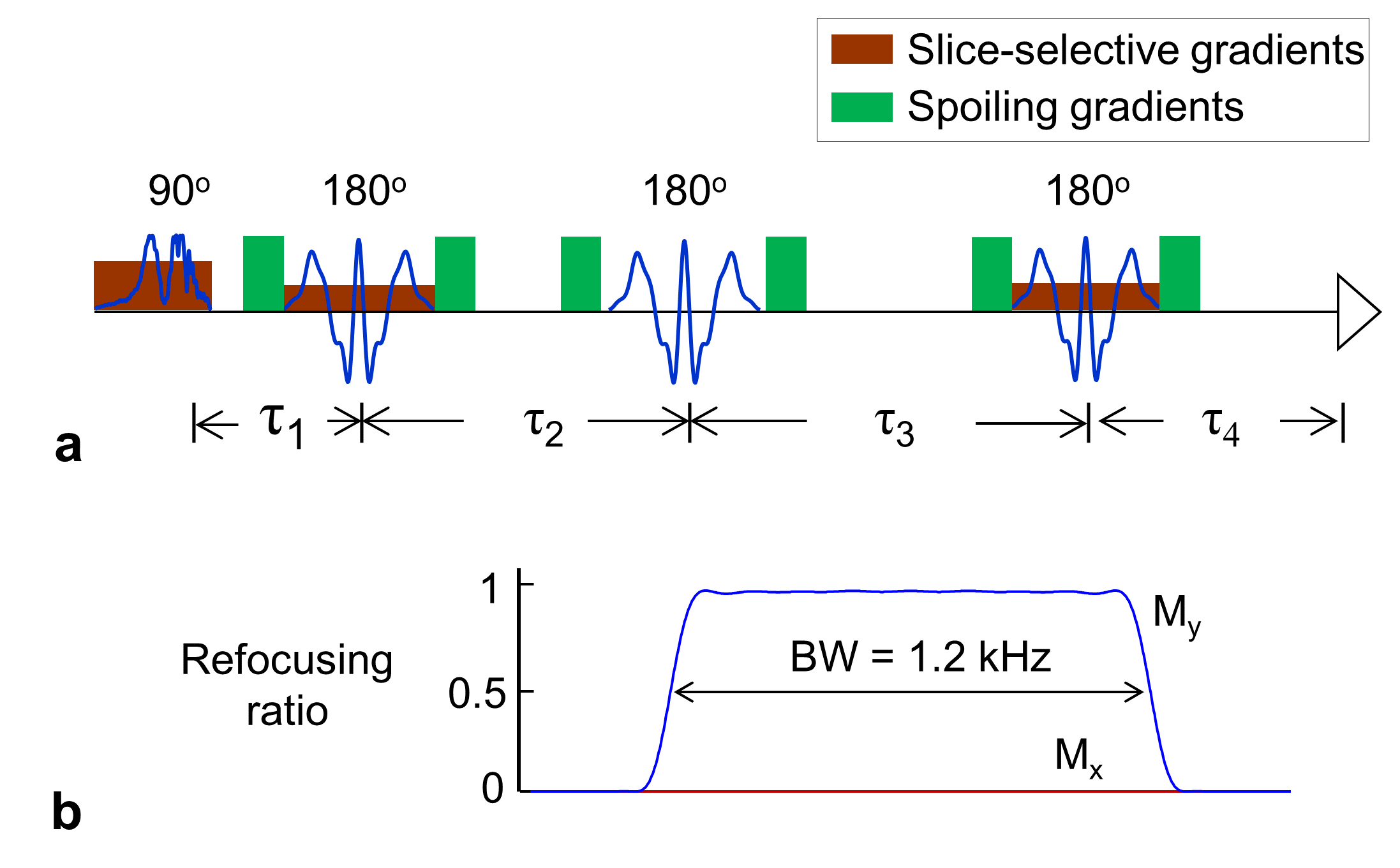
The 1H MRS sequence used had three 180° RF pulses following a slice selective 90° excitation pulse. The first and third 180° pulses were slice selective (13.2 ms; bandwidth 1.3 kHz) and the second 180° pulse was non-slice selective. Volume-localized density-matrix simulations were conducted for optimizing the second 180° pulse duration and the triple-refocusing inter-RF pulse delay times τ1, τ2, τ3 and τ4 (Fig 1.) Restriction of τ1+τ3 = τ2+τ4 for forming an echo and total echo time (TE = τ1+τ2+τ3+τ4) less than 100ms were used for simulation. From ~350,000 calculated spectra, an optimal inter-RF pulse delay time set was obtained with the criteria: 1) largest GABA signal amplitude and most flat Glu signal around 2.29 ppm; 2) small apparent Glu signal linewidth around 2.35 ppm for better separation between Glu and GABA signals.
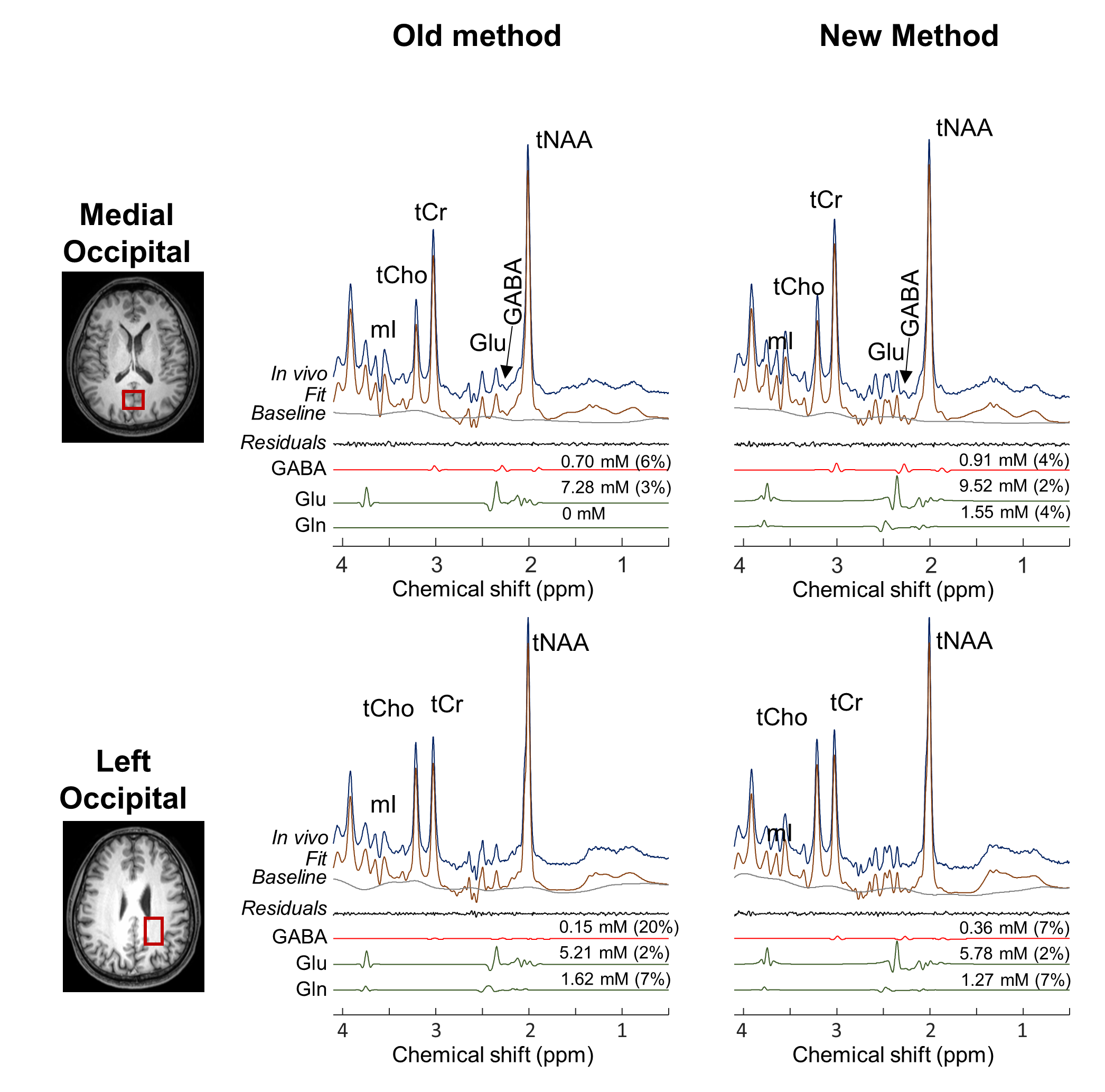
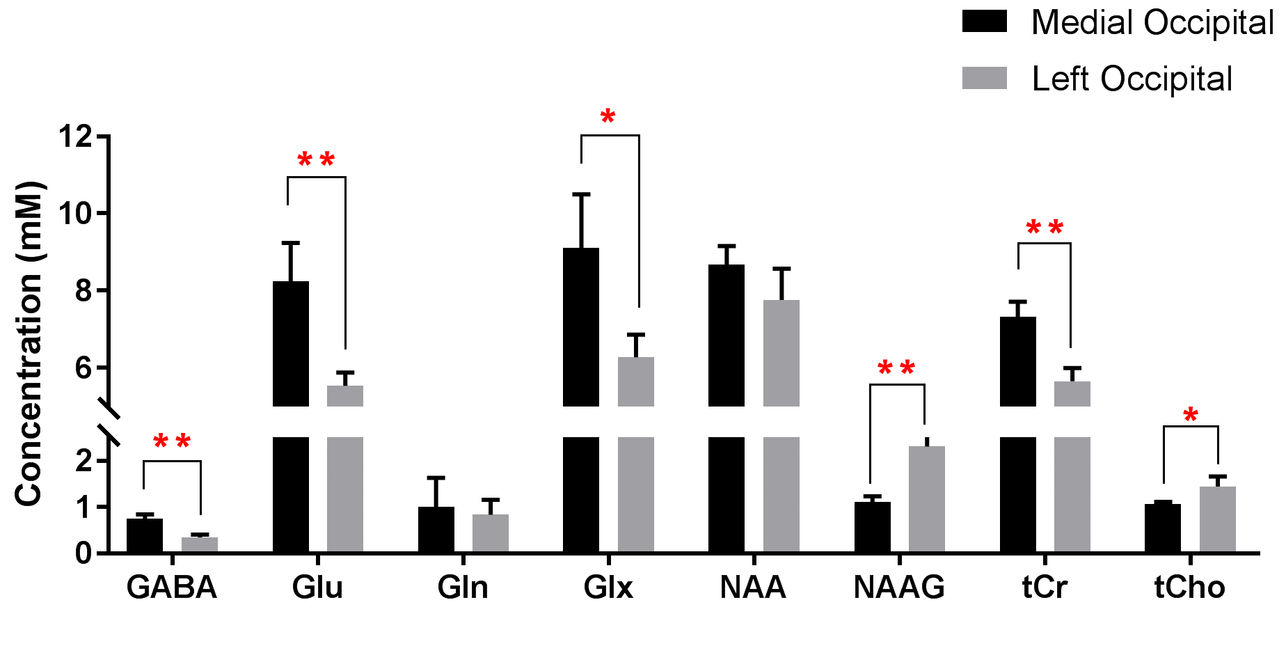
Six healthy adult subjects (3 male and 3 female, age 27±5) were recruited for the study. In vivo 1H MRS data were obtained with proposed triple-refocusing MRS from Medial Occipital (MO) and Left Occipital (LO). The voxel size was 23´23´23 mm3 for MO and 35x23x15 mm3 for LO. Data acquisition parameters included NEX=128 and TR=2 s (scan time 4 min). Data were acquired with a 32-channel head coil in a 3T whole-body scanner (Philips Medical Systems). T1w-MPRAGE was acquired and used for segmentation of gray matter and white matter contents within the voxels. Spectral fitting was performed, with LCModel software,3 using in-house calculated basis spectra of 20 metabolites. Metabolites were quantified with reference to water at 45 M. T2 relaxation effects were corrected using published T2 values.4 Two-tailed paired t-test was performed for each metabolite estimations between MO and LO. Effects for multiple comparisons were corrected by applying Bonferroni correction (p = 0.05/8 = 0.0063).
RESULTS
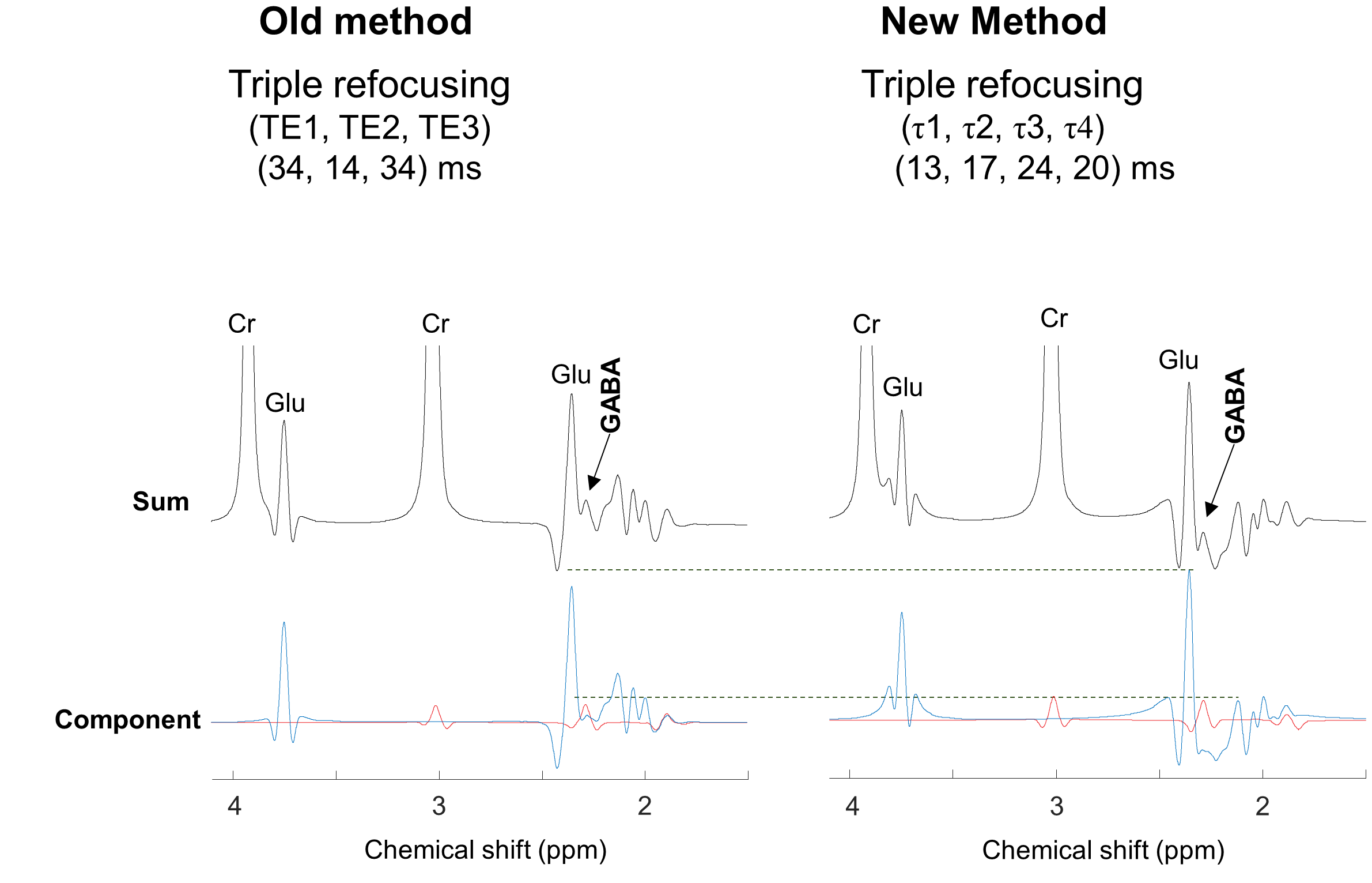
A triple-refocusing subecho time set of inter-RF pulse delay times (τ1, τ2, τ3, τ4) = (13, 17, 24, 20)ms total TE = 74 ms, and a 14ms non-slice selective 180° pulse (1.2kHz bandwidth), tuned to 4.5-ppm was found to have 9.5% higher GABA signal at 2.29ppm and 1Hz narrower and 10% higher Glu signal at 2.35ppm than previously reported subecho time set of (TE1,TE2,TE3) = (34,14,34)ms, total TE = 82ms. Figure 2 shows comparison of numerical simulation results of old method and new method. With 1Hz narrowed Glu signal at 2.35ppm, GABA signal is better differentiable from Glu in newly proposed method. Figure 3 is the representative in vivo spectra of old and new methods acquired from MO and LO of a healthy subject together with LCModel fit and individual metabolite spectra. New method showed an improved detectability and reliability of GABA and Glu with lower CRLBs (6% and 20% vs 4% and 7% for GABA). The new method not only benefits from improved apparent resolution and signal amplitude of GABA and Glu, but also less T2 loss with shorter total TE (74 ms vs 82 ms). GABA was estimated to be 0.91 and 0.36mM for MO and LO, respectively. The mean concentration of GABA was significantly higher in MO than in LO (0.77 vs. 0.35mM; p < 0.001) (Fig.3), in agreement with prior studies.6,7. The mean GABA CRLB was 5% and 9% in MO and LO, respectively, which was significantly improved from old method of 7% and 13%.2 Glu, Glu+Gln (Glx), total Creatine (tCr) and total Choline (tCho) were significantly higher in MO than LO, while N-acetylaspartylglutamate (NAAG) was significantly lower.DISCUSSION AND CONCLUSION
We report a new triple-refocusing MRS method that provides better separation of the GABA 2.29-ppm signal from the Glu C4-proton resonance than the previously reported method. The four-τ scheme provides a wider range for searching optimal sequence parameter which enabled a batter separation and detectability of GABA and Glu with higher signal yield of GABA and narrower Glu C4-proton signal. In conclusion, the newly proposed triple-refocusing method can detect GABA and Glu in the human brain with improved reliability and detectability in a single-shot manner.Acknowledgements
This work was supported by NIH R01CA184584 and CPRIT RP130427.References
1. Tran T, Ross B, Lin A. Magnetic resonance spectroscopy in neurological diagnosis. Neurol Clin. 2008; 27: 21-60.
2. An Z, Ganji SK, Tiwari V, Choi C. Novel Triple-refocusing 1H MRS at 3T for detection of GABA in human brain in vivo. p2370, ISMRM 23rd Annual Meeting & Exhibition, Toronto, Canada, 2015.
3. Provencher SW. Estimation of metabolite concentrations from localized in vivo proton NMR spectra. Magn Res Med. 1993; 30: 672-9.
4. Ganji SK, Banerjee A, Patel AM, et al. T2 measurement of J-coupled metabolites in the human brain at 3T. NMR Biomed. 2012; 25: 523-9.
5. Puts NA, Edden RA. In vivo magnetic resonance spectroscopy of GABA: a methodological review. Prog Nucl Magn Reson Spectrosc. 2012; 60: 29-41
6. Bhattacharyya PK, Phillips MD, Stone LA, Lowe MJ. In vivo magnetic resonance spectroscopy measurement of gray-matter and white-matter gamma-aminobutyric acid concentration in sensorimotor cortex using a motion-controlled MEGA point-resolved spectroscopy sequence. Magn Reson Imaging. 2011; 29: 374-9.
7. Ganji SK, An Z, et al. Measurement of Regional variation of GABA in the human brain by optimized point-resolved spectroscopy at 7T invivo. NMR Biomed. 2014; 27: 1167-1175.
Figures