2977
Evaluation of the amygdala and the anterior cingulate cortex by single voxel proton MR spectroscopy in patients with post-traumatic stress disorder after earthquake1Huaxi MR Research Center, Department of Radiology, West China Hospital of Sichuan University, chengdu, People's Republic of China
Synopsis
In order to explore the changes of metabolites in the amygdala and the anterior cingulate of PTSD after earthquake, MR spectroscopy was applied. MRS showed higher ml and NAA levels in ACC, higher NAA levels in the right amygdala of PTSD group as compared to healthy controls. Besides, both in PTSD and healthy controls, NAA+NAAG levels were higher in the left amygdala than the right. And our results indicate there are some metabolic changes in amygdala and ACC of PTSD subjects. Whether combining depressive disorders, cause of PTSD and types of traumatic causes may have contributed to the inconsistency.
Purpose
Method
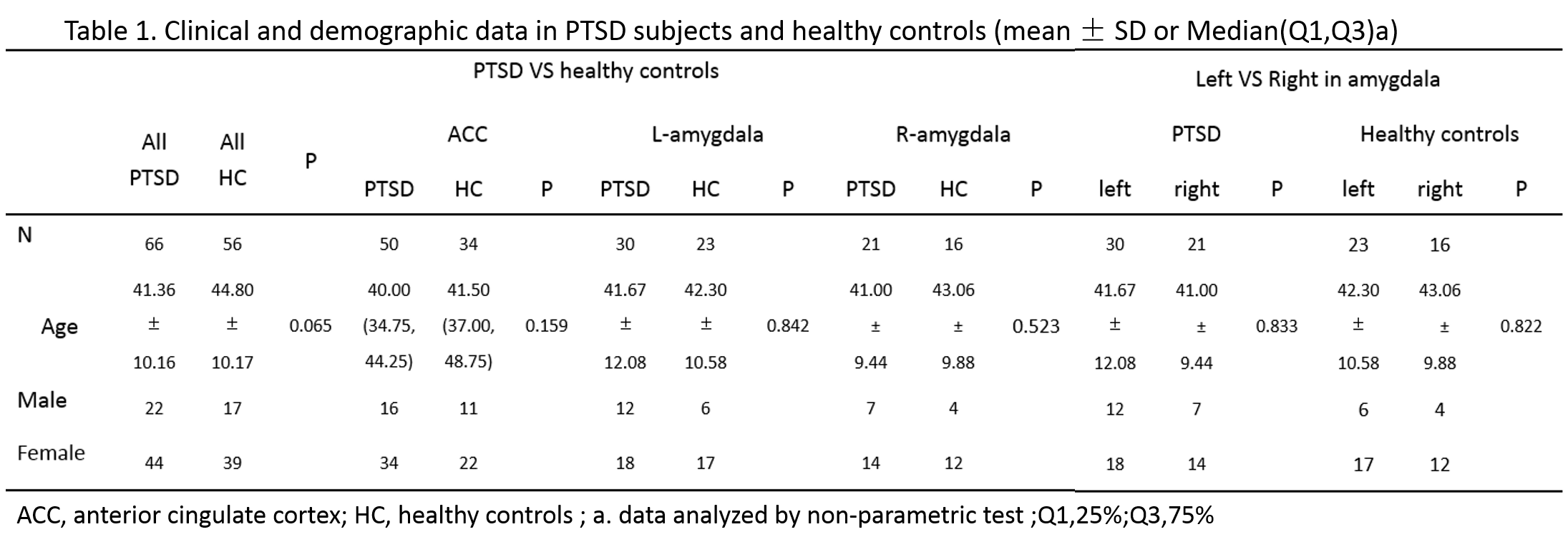
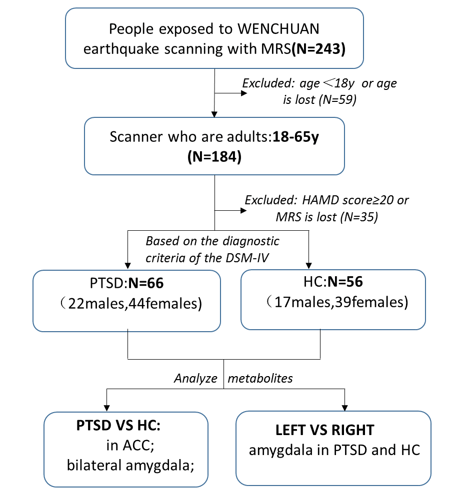
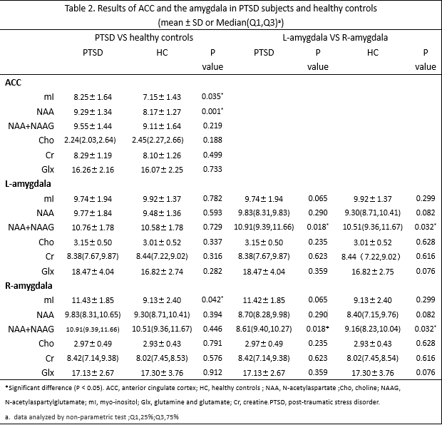
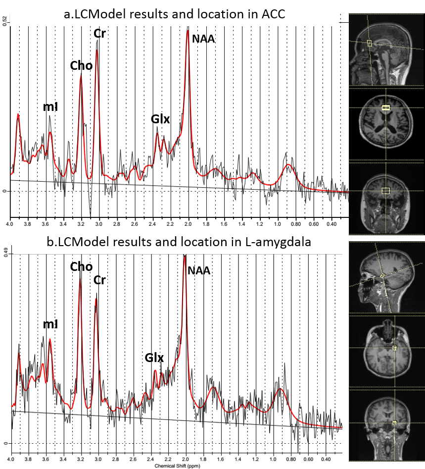
All participants in this study had suffered from a big earthquake in May 2008 in Wenchuan of China. The diagnosis of PTSD were based on Diagnostic and Statistical Manual of Mental Disorders-IV (DSM-IV). 1 Then , depression levels were assessed using the 21-item Hamilton Depression Rating Scale (HAMD) , participants with scores <20 on HAMD-21 were included. All participants were right-hand individuals who were adults, 18 to 65 years old. Exclusionary criteria for all groups included2: a. other axis I psychiatric diagnosis; b. use of psychotropic medications in past 4 weeks; c. any significant medical or neurological conditions or a history of head injury. At last, sixty-six patients with PTSD (22males, 44 females) and fifty-six healthy controls (17males, 39 females) were involved. Age and sex were matched between two groups (Fig 1) . MRS examination was performed on MAGNETOM Germany 3T MR scanner. The normal size of the voxel of interest (VOI) was 20 × 20 × 20 mm3. The data of metabolites were calculated by LCModel, including creatine(Cr),N-acetylaspartate (NAA), NAA+N-acetylaspartylglutamate(NAAG), choline (Cho), myo-inositol (mI),glutamine and glutamate(Glx). The low-quality MRS data were excluded (Cr SNR<15, FWHM >0.08ppm). First, we compared metabolites of anterior cingulate cortex (ACC) and bilateral amygdala between PTSD and HC. Then, respectively compared the left and right amygdala in PTSD subjects and HC (Fig 2) .Result
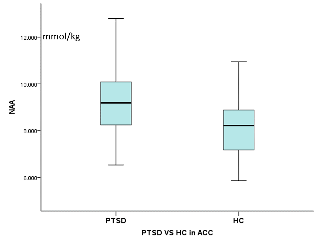
As compared to healthy controls, analysis of the proton MRS showed higher mI levels (P=0.035, 28PTSD VS 15HC) and higher NAA levels (P=0.001,42PTSD VS 29HC) in ACC, higher NAA levels(P=0.042,9PTSD VS 8HC) in the right amygdala of PTSD subjects .When compared the left and right of amygdala, MRS showed significantly higher NAA+NAAG levels in the left amygdala of both group (PTSD:P=0.018, 26left VS 21right;HC:P=0.032,21left VS 13right). (Fig 3) .Discussion and Conclusion
Acknowledgements
No acknowledgement found.References
1. Amerikan Psikiyatri Birligi: Mental Bozukluklarin Tanisal ve Sayimsal El Kitabi,4. baski (DSM IV), Amerikan Psikiyatri Birligi, Washington DC, 1994. Köroglu E, çeviri ed. Hekimler Yayin Birligi, Ankara, 1994; 179-194.
2. ZhiYong Yang, Hong Quan, ZuLai Peng, et al. Proton magnetic resonance spectroscopy revealed differences in the glutamate + glutamine/creatine ratio of the anterior cingulate cortex between healthy and pediatric post-traumatic stress disorder patients diagnosed after 2008 Wenchuan earthquake. Psychiatry and
3. Rauch SL, Shin LM, Phelps EA. Neurocircuitry models of posttraumatic stress disorder and extinction: Human neuroimaging research – Past, present, and future. Biol. Psychiatry .2006; 60: 376–382. Clinical Neurosciences .2015; 69: 782–790.
4. Byung-Joo Ham, Jeanyung Chey, Sujung J. Yoon,et al. Decreased N-acetyl-aspartate levels in anterior cingulate and hippocampus in subjects with post-traumatic stress disorder: a proton magnetic resonance spectroscopy study. European Journal of Neuroscience. 2007,25: 324–329.
5. Norbert Schuffa, Thomas C. Neylanb, Sabrina Fox-Bosettia,et,al. Psychiatry Research:Neuroimaging. 2008, 162:147–157.
6. Griffin JL, Bollard M, Nicholson JK, et al. Spectral profiles of cultured neuronal and glial cells derived from HRMAS (1)H NMR spectroscopy. NMR Biomed ,2002,15: 375–384.
7. Chen LP, Dai HY, Dai ZZ, et al. Anterior cingulate cortex and cerebellar hemisphere neurometabolite changes in depression treatment: A 1H magnetic resonance spectroscopy study. Psychiatry Clin Neurosci .2014,68: 357–364.
8. Davis M. The role of the amygdala in fear and anxiety. Annu Rev Neurosci.1992,15:353-375.
9. Davis M. The role of the amygdala in fear-potentiated startle: implications for animal models of anxiety. Trends Pharmacol Sci. 1992,13:35-41.
10. Jayasundar R. Human brain: biochemical lateralization in normal subjects. Neurol India.2002;50:267- 71.
11. G. Castellano, C.S.B. Dias, B. Foerster, et al. NAA and NAAG variation in neuronal activation during visual stimulation. Brazilian Journal of Medical and Biological Research.2012,45: 1031-1036.
Figures