2959
In Vivo Double Quantum Filtered 23Na Imaging of Human Skeletal Muscle1Institute of Radiology, University Hospital Erlangen, Erlangen, Germany
Synopsis
Double quantum filtered sodium (23Na) MRI represents a way to examine the degree of tissue microstructure but suffers from low signal intensity and therefore long acquisition time. In this work we developed an efficient simultaneous acquisition scheme of single and double quantum images and determined the optimum acquisition parameters for human skeletal muscle. The feasibility of double quantum filtered 23Na images of human lower leg at 3T was shown.
Purpose
Over the past years 23Na MRI has become a widely used technique to noninvasively determine the total tissue sodium concentration or even to distinguish intracellular and extracellular sodium. However, its full potential is still far from being exploited as it also offers the possibility to gain information about the sodium ions’ molecular environment. Specific detection of sodium ions within ordered structures can be realized using double quantum filtered 23Na MRI.1 A change in order may represent an additional indicator for certain pathologies. So far only human brain has been examined using double quantum filtered sodium imaging2 but also skeletal muscle is expected to generate a sodium DQ signal.3 This assumption is encouraged by recently published double quantum filtered 39K images of human thigh.4 In this work the first double quantum filtered 23Na images of human lower leg have been acquired using a modified double quantum filtered magic angle sequence (DQ-MA) with integrated single quantum (SQ) acquisition.Methods
Pulse sequences were implemented on a 3-Tesla whole-body MR system (Magnetom Skyra, Siemens, Erlangen, Germany). A single-resonant 23Na birdcage coil (Stark Contrast, Erlangen, Germany) was used for the measurements. The total double quantum filtered signal of human lower leg muscles was acquired using a DQ-MA FID sequence to determine the optimum acquisition parameters for double quantum filtered images. The sequence diagram of the DQ-MA imaging sequence is shown in Fig. 1. The acquisition of a spin density weighted sodium image was included in the common DQ-MA sequence between the first and the second RF pulse to enhance efficiency, as proposed for triple quantum filtered measurements.5 To detect the spin density weighted sodium signal a three-dimensional density adapted radial acquisition (DA-3DPR)6 with ultrashort echo time was used. For the acquisition of the double quantum filtered signal a conventional 3D gradient echo (3D-PE) readout was applied. The magic angle of 57.4° was chosen for both the second and third RF pulse to suppress unwanted signal of third-rank double quantum coherences.
Sequence parameters: DQ-MA FID: TR = 200 ms, TE = 0.2 ms, TRO = 20 ms, τ = 1 - 8 ms, δ = 40 µs,100 averages, TA = 10:40 min; SQ/DQ-MA combined: TR = 100 ms, TE,1 = 0.2 ms, TE,2 = 5 ms, TRO,1 = 3 ms, TRO,2 = 8 ms, τ = 4.5 ms, δ = 40 µs, (Δx1)3 = (5mm)3, (Δx2)3 = (15 mm)3, 6 averages, TA = 16:00 min
Results and Discussion
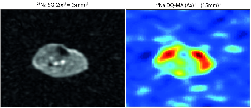
Double quantum filtered sodium FID measurements of the lower leg were conducted at three healthy volunteers. As the individual data points were subject to high fluctuations the total signal intensity integrated over the acquisition time was used for further considerations. The resulting relation between total DQ-MA signal intensity and preparation time τ is shown in Fig. 2 a). All three curves point out a maximum at about τ = 5 ms and a rather broad range of preparation times resulting in high signal intensity (τ = 4 ms - 7ms). The corresponding FIDs measured at τ = 5 ms are shown in Fig. 2b). Additionally, a fit of the expected DQ-MA signal equation in biological tissue7 to the individual FID curves was performed to estimate the fast component of transverse relaxation time (T2f) in muscle. The fit values of the three data sets lead to a mean value of T2f = 4.37 ms with a standard deviation of ΔT2f = 0.25 ms. Fig. 3 compares a single quantum 23Na image of human lower leg with the corresponding double quantum image generated with the combined SQ/DQ-MA acquisition sequence. Using optimized acquisition parameters an isotropic nominal resolution of (15mm)3 for the DQ-MA image and (5mm)3 for the SQ image could be achieved within an acquisition time of TA = 16:00 min. As expected due to muscle fibers' structure, significant double quantum filtered signal can be found in both calf muscles and tibialis anterior muscle. Due to low resolution and resulting partial volume effects no further compartments within the muscles can be distinguished.
Conclusion
In this work the first in vivo double quantum filtered 23Na images of human lower leg have been presented. For this purpose, an efficient combined single and double quantum acquisition scheme was developed and the optimum preparation time was determined. It was shown that a simultaneous acquisition of SQ and DQ-MA 23Na images is feasible at 3T within reasonable acquisition time using the described imaging sequence.Acknowledgements
No acknowledgement found.References
1. Jaccard G, et al. Multiple-quantum NMR spectroscopy of S=3/2 spins in isotropic phase: A new probe for multiexponential relaxation. Journal of Chemical Physics 85 (11):6282-6293, 1986.
2. Tsang A, et al. In vivo double quantum filtered sodium magnetic resonance imaging of human brain. Magnetic Resonance in Medicine, 73(2):497-504, 2015.
3. Reddy R, et al. Detection of residual quadrupolar interaction in human skeletal muscle and brain in vivo via multiple quantum filtered sodium NMR spectra. Magnetic Resonance in Medicine, 33(1):134-139, 1995.
4. Rösler MB, et al. In vivo observation of quadrupolar splitting in 39K magnetic resonance spectroscopy of human muscle tissue. NMR in Biomedicine, 29:451-457, 2016.
5. Fiege DP, et al. Simultaneous single-quantum and triple-quantum-filtered MRI of 23Na (SISTINA). Magnetic Resonance in Medicine, 69(6):1691-1696, 2013.
6. Nagel AM, et al. Sodium MRI using a density-adapted 3D radial acquisition technique. Magnetic Resonance in Medicine, 62(6):1565-1574, 2009.
7. Hancu I, et al. A model for the dynamics of spins 3/2 in biological media: signal loss during radio-frequency excitation in triple-quantum filtered sodium MRI. Journal Of Magnetic Resonance, 147(2):149-191, 2000.
Figures
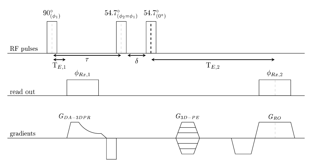
Fig. 1: Sequence diagram of combined SQ and DQ-MA acquisition. A four step phase cycling scheme (Φ1= Φ2 = 0°, 90°, 180°, 270°) and ΦRx,2 alternating between 0° and 180° was applied. The phase of the first ADC was set to match the phase of the first RF pulse. The evolution time δ was chosen as short as possible (40 μs).
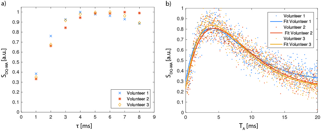
Fig. 2: a) Total time integrated 23Na DQ-MA signal intensity of lower leg depending on preparation time. Three healthy volunteers were scanned. b) DQ-MA FIDs measured at τ = 5 ms with fits of theoretically expected signal curves.