2958
Elliptically-shaped 1Tx4Rx Coil for 23Na Body MRI at 7T1Medical Physics in Radiology, German Cancer Research Center (DKFZ), Heidelberg, Germany, 2Institute of Radiology, University Hospital Erlangen, Erlangen, Germany, 3Diagnostic and Interventional Radiology, University Hospital Heidelberg, Heidelberg, Germany
Synopsis
Up to now, only a few abdominal 23Na-MRI studies have been performed at 7T. In this work a 23Na body coil for 7T was enhanced. To achieve an improved homogeneity in the transmit field and in the receive sensitivity, transmit phase settings were optimized and four separate receive channels were implemented. Field distributions in a phantom were obtained for the original and the enhanced configuration. Both setups were applied to the human chest and abdomen. Transmit and receive homogeneity are markedly improved for the enhanced setup. Improvements for in-vivo image quality are especially visible in the contour of the body.
Purpose
Over the last decades, sodium (23Na) MRI has evolved into a valuable biomedical research tool which can give additional information about tissue viability. However, 23Na-MRI is challenging due to the inherently low signal-to-noise ratio (SNR). Up to now, only a few 23Na abdominal MRI studies have been performed at 7T, mainly due to the lack of availability of 23Na body coils1. In this work a versatile 23Na body coil for 7T was enhanced by extending the original setup2. To achieve an improved homogeneity in the transmit field and in the receive sensitivity, transmit phase settings were optimized and four separate receive channels (1Tx4Rx) were implemented. To demonstrate this, maps of relative flip angle and receive sensitivity inside a phantom were obtained from RF simulations and measurements. Furthermore, both hardware configurations (1Tx1Rx, 1Tx4Rx) were applied for in-vivo 23Na imaging of the human chest and the human abdomen.
Methods
Coil:2 A close-fitting low-pass elliptical birdcage was used (f23Na=78.6MHz). Due to the violated rotational symmetry of the elliptical geometry, the generation of a circularly-polarized B1+-field is challenging. To improve quadrature performance the legs were spaced 360°/12=30° apart around the periphery of the ellipse3, and to improve B1+-homogeneity four feeding ports were implemented4. The left and right legs of the birdcage had to be shifted to the upper half (Fig.1a), resulting in an additional violation of symmetry.
Hardware development: For the original 1Tx1Rx configuration2 (Fig.1a,b), four quadrature hybrids provide phase differences of 0°, -90°, -180°, -270° for transmission and, moreover, combine the four receive signals. For the enhanced 1Tx4Rx configuration (Fig.1c,d), phase shifts were optimized in RF simulations to achieve a symmetric transversal B1+-field with respect to the coil center. Resulting phase differences of -17°, -124°, -197°, -270° were generated with four quadrature hybrids and additional phase shifts. The four receive signals were amplified and were directed to the system receivers. For this purpose four transmit/receive switches were required instead of one as for 1Tx1Rx.
Coil simulations: RF simulations for both configurations (1Tx1Rx, 1Tx4Rx) were performed in CST5. B1+-maps and relative receive sensitivity maps were calculated for a phantom. Relative flip angle maps were determined from B1+-maps. For 1Tx1Rx, receive signals were combined to one signal according to the RF hardware. For 1Tx4Rx, receive sensitivity was determined from a sum-of-squares (SOS) combination of the four receive signals. Homogeneity in the transmit field and in the receive sensitivity was investigated by evaluating the coefficient of variation (CV).
Imaging: Measurements were performed on a 7T whole-body MR system6. 23Na images were acquired using a 3D density-adapted radial sequence7. For 1Tx4Rx, a SOS combination was performed.
Relative flip angle maps inside a phantom were obtained using the dual-angle method8. Relative receive sensitivity maps were determined from the same data. Homogeneity of these distributions was investigated by evaluating the CV.
3D in-vivo 23Na-MRI of the human chest and the human abdomen of a free-breathing healthy volunteer (female, 27y/o) were obtained with 1Tx1Rx configuration and with 1Tx4Rx configuration. SNR was compared for the heart. Sequence parameters: Nominal resolution=(4mm)3, FA=44°, tp=2ms, TE/TR=1.05ms/20ms, Nprojections=18200, avg=5, tacq=30min20s. Reconstruction: zero-filling factor=2, Hamming filter, reconstructed FOV=(400mm)3.
Results
Phantom study: Simulated and measured maps of transmit field (Fig.2, Table) correspond very well for both hardware configurations. As desired, the transversal field distribution for optimized transmit phases (1Tx4Rx) is approximately symmetric with respect to the coil center. For 1Tx4Rx, the measured CV is reduced by a factor of approx. 0.6 (Δz=10cm) and approx. 0.7 (Δz=20cm) compared to 1Tx1Rx.
Simulated and measured distribution of receive sensitivity (Fig.3, Table) also correspond very well for both configurations. For 1Tx4Rx, the measured CV is reduced by a factor of approx. 0.4 (Δz=10cm) and approx. 0.7 (Δz=20cm) compared to 1Tx1Rx.
In-vivo study: SNR was improved for 1Tx4Rx especially for 23Na-MRI of the human chest (Fig.4). SNR of the heart was increased in the delineated ROI (Fig.4) by approx. 40%. Improvements in image quality are especially visible for the contour of the body (white arrows).
Discussion & Conclusion
Transmit and receive homogeneity are markedly improved for the enhanced configuration, which provides four receive signals (1Tx4Rx) instead of one (1Tx1Rx). In the present work a conventional SOS combination was applied. However, to exploit the capabilities of four transmit channels, reconstruction could be performed using adaptive combination9,10.
The developed body coil offers the possibility to examine in-vivo 23Na distribution at 7T in organs such as heart, breast, lung, kidney, and liver. Furthermore, the presented hardware extension provides an improved in-vivo 23Na image quality especially in tissues next to the contour of the body such as skin and breast.
Acknowledgements
This work was funded in part by the Helmholtz Alliance ICEMED - Imaging and Curing Environmental Metabolic Diseases, through the Initiative and Networking Fund of the Helmholtz Association.
This study was also supported by grants from the Bundesministerium für Bildung und Forschung (BMBF) to the German Center for Lung Research (DZL) (82DZL00401 and 82DZL004A1).
References
1) Madelin, G. & Regatte, R.R. J Magn Reson Imaging (2013) 38: 511–29
2) Platt, T. et al. Proc Intl Soc Mag Reson Med (2016) 24: 3977
3) Kurczewski, R. et al. Proc Intl Soc Mag Reson Med (1992): 4025
4) Ibrahim, T.S. et al. Magn Reson Imag (2000) 18.6: 733-42
5) CST Studio Suite 2016, CST AG, Darmstadt, Germany
6) MAGNETOM 7T, Siemens Healthcare, Erlangen, Germany
7) Nagel, A.M. et al. Magn Reson Med (2009) 62: 1565-73
8) Insko, E.K. & Bolinger, L. Journal of Magnetic Resonance (1993) Series A, 103: 82
9) Walsh, D.O. et al. Magn Reson Med (2000) 43: 682-90
10) Benkhedah, N. et al. Magn Reson Med (2016) 75(2): 527-36
Figures
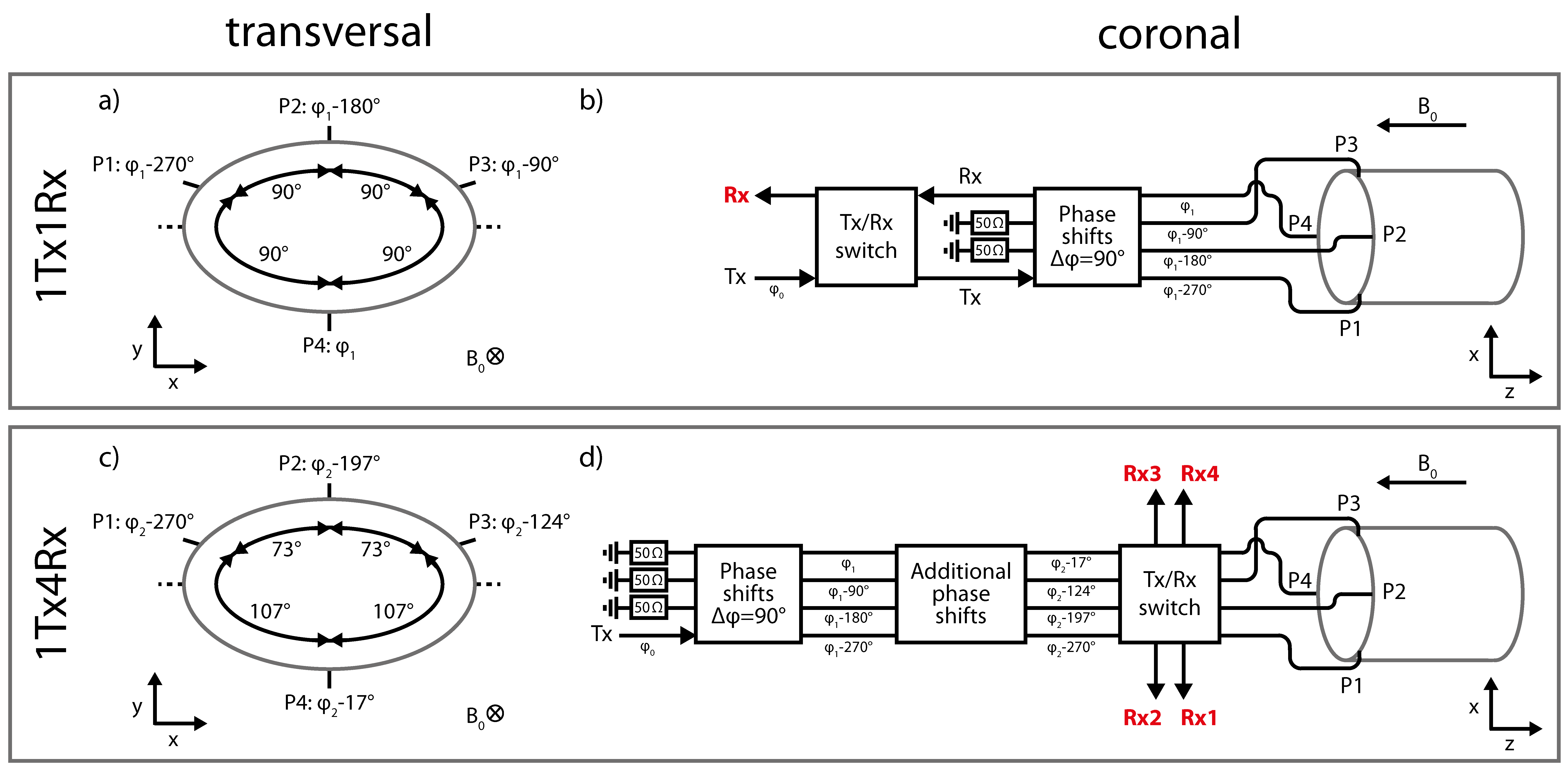
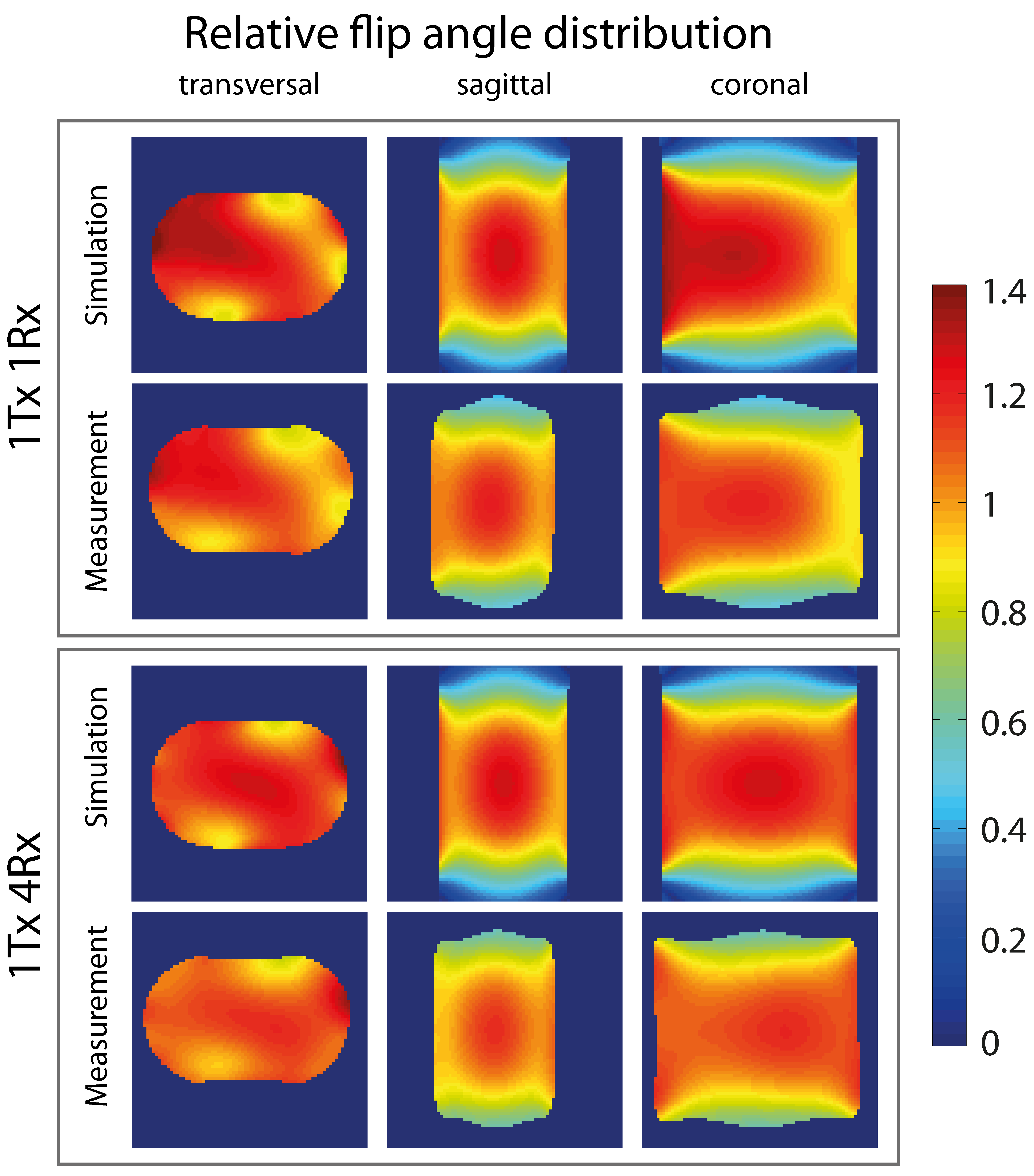
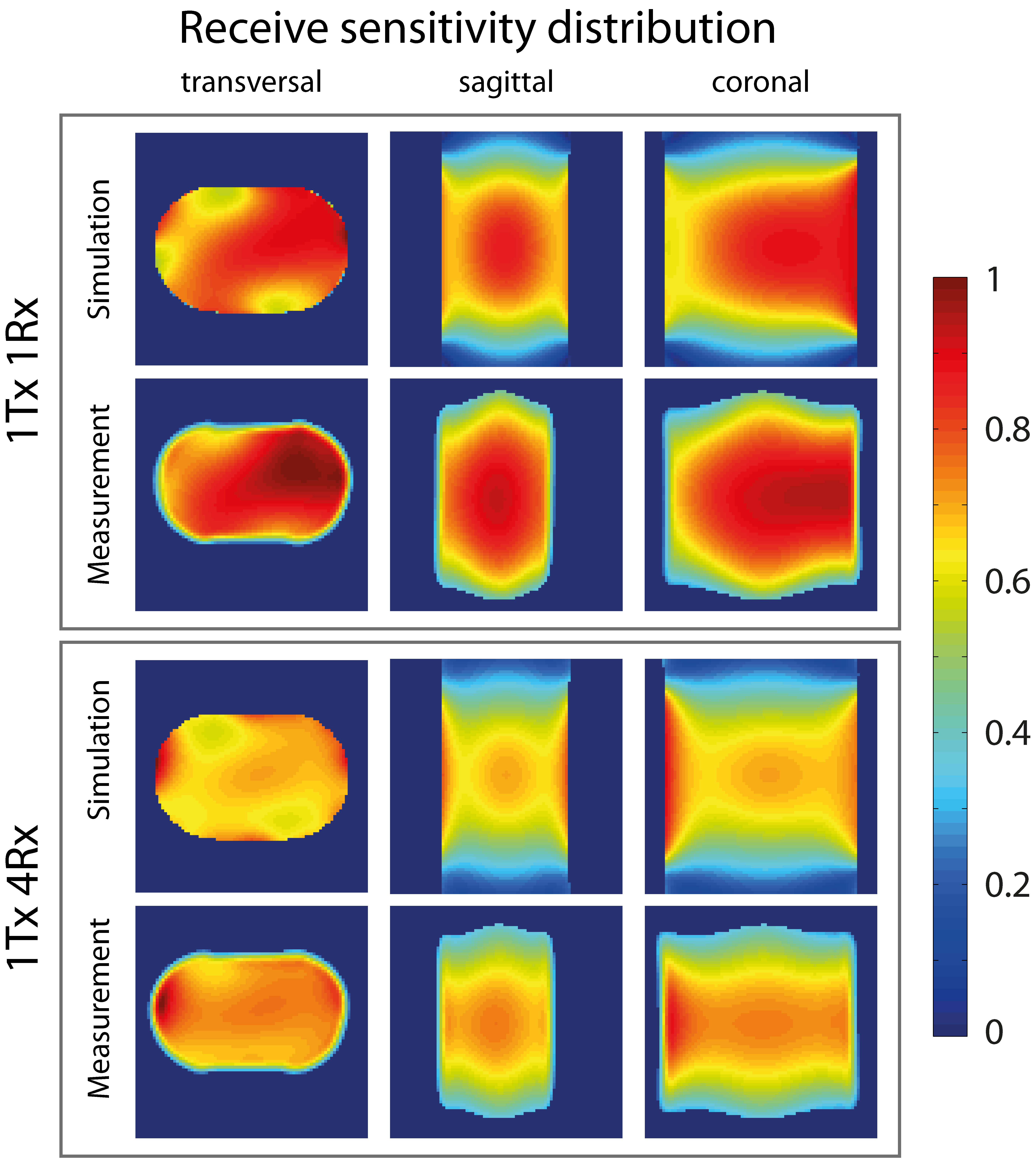
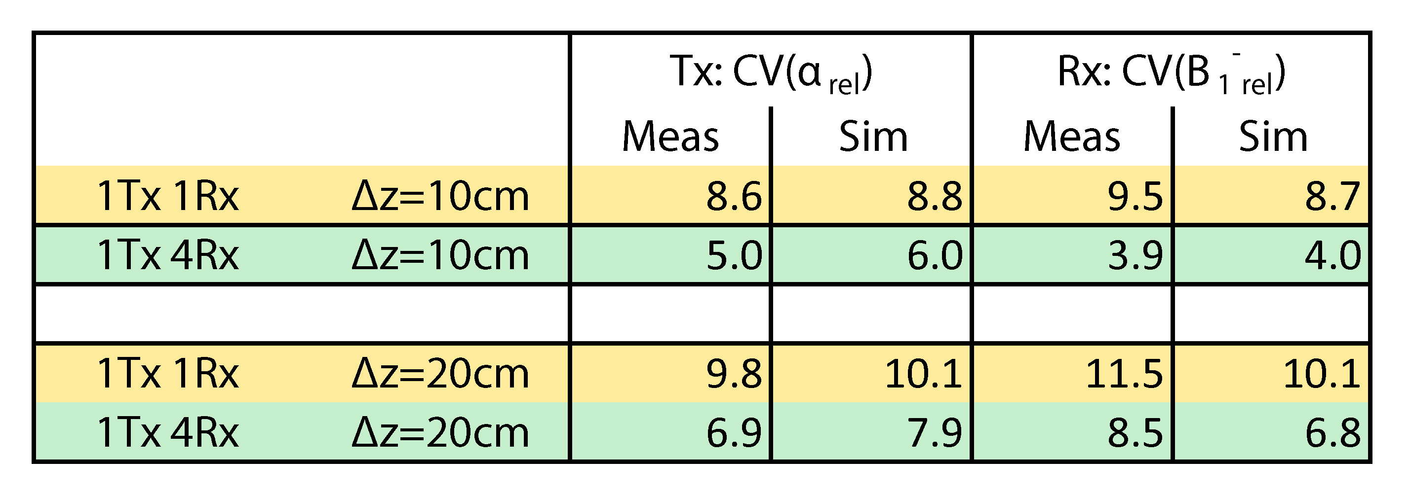
Fig.4: Comparison of in-vivo 23Na-MRI for the original configuration (top) and for the enhanced configuration (bottom) at B0=7T and with tacq(per dataset)=30min20s. SNR is improved for 1Tx4Rx especially for the chest (left). SNR of the heart is increased by approx. 40% (ROI). Improvements in image quality are visible for the contour of the body (white arrows). Tissues such as skin and breast can only be reliably detected with 1Tx4Rx using these imaging parameters. In chest MRI the heart is particularly prominent, whereas in abdominal MRI the kidneys are prominent. Further signals arise from liver, stomach, cartilage, and intervertebral disks.