2949
Density-Adapted k-Space Sampling Technique for Fast Relaxing Chlorine-35 Nuclei at 9.4 T1Computer Assisted Clinical Medicine, Heidelberg University, Mannheim, Germany, 2MR-Imaging and Spectroscopy, Universty of Bremen, Bremen, Germany
Synopsis
Chloride Cl- as the most abundant anion in mammals is a vital component of biophysical regulatory processes. 35Cl MRI has the potential to visualise Cl- homeostatic changes during stroke, in tumors and in ionic regulatory diseases. This study aimed at demonstrating the advantages of implementing density-adapted k-space sampling to overcome the instrinsically low SNR associated with 35Cl MRI. We showed that density-adapted k-space sampling yielded distinctively visible improvement of image quality as well as quantitative SNR gain in phantoms with very fast bi-exponential T2* relaxation over conventional radial sampling.
Purpose
Chloride Cl- as the most abundant anion in mammals is a vital component of biophysical regulatory processes. 35Cl MRI (spin = 3/2) has the potential to visualise Cl- homeostatic changes during stroke1, in tumors and in ionic regulatory diseases2. The low gyromagnetic ratio, low biological concentration and fast transversal relaxation result in an extremely low intrinsic signal-to-noise ratio (SNR). One approach to overcome the SNR-challenge is offered by density-adapted 3D projection reconstruction3 (DA-3DPR), which has been implemented for the first time on a 9.4 T preclinical system to the best of our knowledge. It is a center-out non-Cartesian imaging technique whose gradients are designed such that the averaged sampling density in each spherical shell of k-space remains constant. DA-3DPR enables ultrashort echo times, benefits from the straightforward k-space trajectory design of conventional radial sampling techniques and furthermore achieves the same intrinsic SNR efficiency as twisted projection imaging4,5.
This study aimed at investigating the advantages of implementing density-adapted k-space sampling provided by DA-3DPR to overcome the low SNR challenges especially associated with 35Cl MRI in environments with very fast bi-exponential T2* relaxation.
Materials and Methods
Hardware and phantoms: Experiments were performed on a 9.4 T preclinical MR system (BioSpec, Bruker, Germany) using a custom-made 16 rung low-pass birdcage 35Cl volume coil. Three phantoms in 50 ml vials with 134.75 mM NaCl and different agarose concentrations (2, 4 and 6 %) were used.
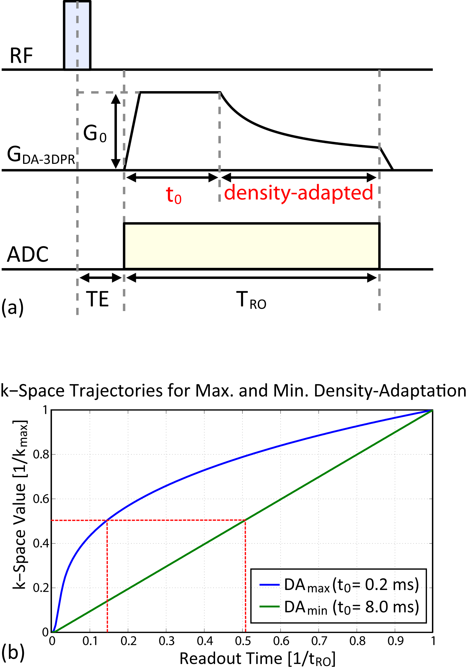
DA-3DPR properties: The parameter t0 in the gradient design (cf. Figure 1a) is the time between the beginning of the gradient and the beginning of the density-adapted section. By varying t0 the percentage of the density-adapted gradient section is altered. The theoretically maximum percentage of density-adaption (DAmax,theo) for t0 = 0 ms is unachievable due to hardware limitation; for t0 = tRO (DAmin) the gradient takes the conventional trapezoidal shape, thus eliminating density-adaptation.
DA-3DPR parameters: Sequence parameters were: readout bandwidth = 25 kHz, 200 sampling points per projection, tRO = 8 ms, TE = 306 µs, TR = 80 ms, α = 80°, tRF = 600 µs, 8 averages. Two series of experiments were conducted at varying isotropic resolution (RES):
RES1.96mm: FOV = (70.56 mm)³, projection = 4000, tscan = 42:40 min, t0 varied between 0.5–8.0 ms (corresponded to DAmax–DAmin), G0 varied between 36.20–7.66 mT/m.
RES3.92mm: FOV = (78.40 mm)³, projection = 1000, tscan = 10:40 min, t0 varied between 0.2–8.0 ms (corresponded to DAmax–DAmin), G0 varied between 36.54–3.83 mT/m.
Exemplatory k-space trajectories for t0 = 0.2 and 8.0 ms are shown in Figure 1b.
Results and Discussion
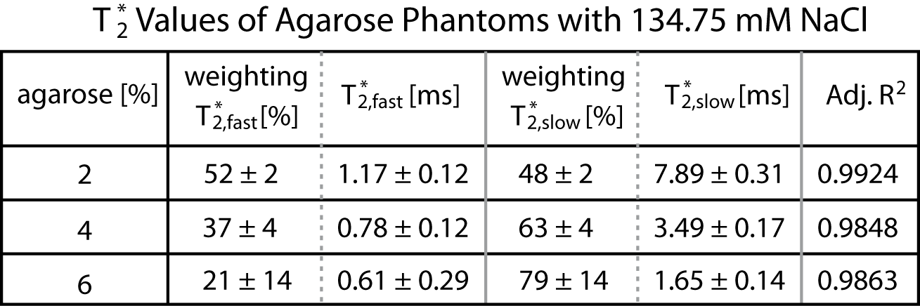
T2* measurements: The phantoms share the same range of T2* (cf. Figure 2) with rat brain tissues1. T2,fast* values lie below 1.5 ms, emphasizing the neccessity of efficient k-space sampling immediately after RF excitation.
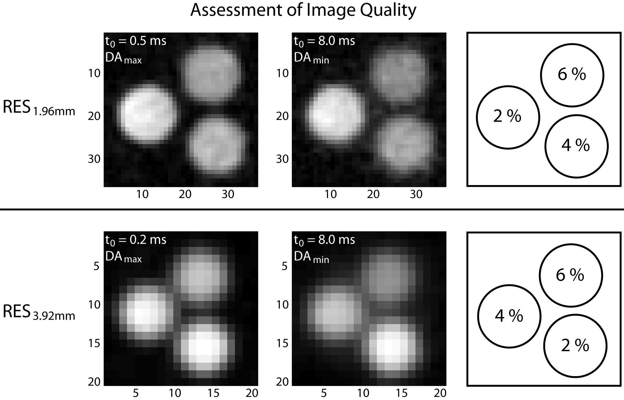
Assessment of image quality: Phantom images (cf. Figure 3) show sharper edges and a more homogeneous background at DAmax than those at DAmin. Phantoms with a higher agarose concentration exhibit more severe signal loss at DAmin. Uneven smearing of the phantom border is clearly visible in images acquired with RES1.96mm at DAmax. For images acquired with RES3.92mm, partial volume effects negatively influence the differentiation between neighbouring phantoms more gravely at DAmin than at DAmax.
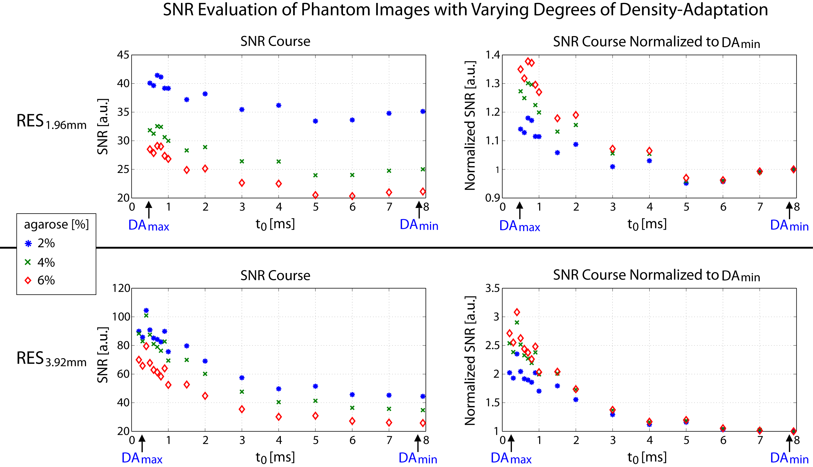
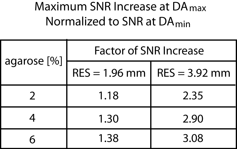
Quantitative assessment via SNR calculation: SNR of each phantom is calculated from the images via $$\small\tt SNR=\frac{mean(ROI_{phantom})-mean(ROI_{background})}{std(ROI_{background})}.$$ SNR of each phantom acquired with different t0 were normalized to that with t0 = tRO (cf. Figure 4). Without normalization, phantoms with lower agarose content have higher SNR because of the faster T2* relaxation during the RF excitation. Normalized SNR courses demonstrate that the faster the T2* relaxation is, the more benefits of using density-adapted gradients are evident. The factor of maximum SNR increase (cf. Figure 5) lies between 1.18–1.38 and 2.35–3.08 in images acquired with RES1.96mm and RES3.92mm, respectively.
Conclusion
Comparison between images with density-adapted k-space sampling and those with conventional gradients show distinctively visible qualitative improvement of the former over the latter especially for phantoms with higher agarose concentration. Quantitative SNR evaluation demonstrates a steady SNR increase with increasing degree of density-adaptation. Furthermore, environments with faster T2* relaxation suffer from more severe signal loss during RF excitation and are in particular need of efficient coverage of the k-space center. Simultaneously, they have been shown to have attained the highest SNR gain provided by density-adaptation.Acknowledgements
No acknowledgement found.References
1. Baier et al. Chlorine and Sodium Chemical Shift Imaging during Acute Stroke in a Rat Model at 9.4 Tesla. MAGMA. 2014;27(1):71-79
2. Nagel et al. In Vivo 35Cl MR Imaging in Humans: A Feasibility Study. Radiology. 2014;271(2):585-595
3. Nagel et al. Sodium MRI Using a Density-Adapted 3D Radial Acquisition Technique. MRM. 2009;62(6):1565-1573
4. Konstandin & Nagel. Measurement Techniques for Magnetic Resonance Imaging of Fast Relaxing Nuclei. MAGMA. 2014;27(1):5-19
5. Boada et al. Fast Three Dimensional Sodium Imaging. MRM. 1997;37:706-715
Figures