2929
Multi-parametric MRI of glioblastoma invasion quantitative evaluation using histological stacks1Glasgow Experimental MRI center, University of Glasgow, Glasgow, United Kingdom, 2Institute of Cancer Sciences, University of Glasgow, Glasgow, United Kingdom, 3School of medicine, dentistry and nursing, University of Glasgow, Glasgow, United Kingdom
Synopsis
We perform a quantitative histological evaluation of a range of MRI techniques in their ability to probe glioblastoma invasion in a mouse model. Using 3-D histological stacks co-registered with MRI slices allows to achieve high values in Dice, sensitivity and specificity tests (>90%). This approach enables to go beyond the standard evaluation tests, performing direct voxel-to-voxel comparison between MRI and histology, and facilitating the development of multi-parametric analysis models. We also identified promising methods for detecting low tumour concentration regions at the invasion limits.
Introduction
Magnetic Resonance Imaging (MRI) is an important tool
for glioblastoma (GBM) diagnosis, however, several works have reported limitations
of diagnostic accuracy1 for tumour invasion. In
order to more robustly evaluate MRI techniques, we have developed a protocol for the co-registration of histological
stacks with MRI slices. Care was taken to cut the histological slices (HLA
stain for human GBM) in exactly the
same plane as the MR images, with five evenly distributed 20 mm histological slices then averaged to account for the thicker MRI
slice (1.5mm). We have used this protocol to evaluate the ability of a range of
MRI techniques to probe invasion in a mouse glioblastoma model. This approach allows us to go beyond the standard
evaluation tests and enables a direct voxel-to-voxel comparison between MRI and
histology. Methods
This study used ten nude
mice injected intra-cranially with human glioblastoma2, presenting highly invasive tumour margins. The mice
brains were scanned in vivo and sacrificed at week 12 post-injection. Imaging
was performed on a Bruker 7T Biospec with 72 cm volume resonance and 4 channel phase-array
surface coil. Several imaging modalities
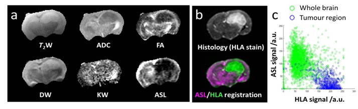
were acquired as following: (Figure 1a): T2 weighted (T2W),
T2 value (T2map), Diffusion weighted (DW), Apparent
Diffusion Coefficient (ADC), Fractional anisotropy (FA), Kurtosis weighted (KW)
and perfusion weighted (ASL). Slice thickness was 1.5 mm and resolution varied
according to the MRI technique used. Five evenly distributed 20 mm histological slices (HLA stain for human GBM) were cut in the MRI
plane (using high resolution T2 images for reference) and stacked to
account for MRI slice thickness. Post processing of the data (with in-house
developed MATLAB code) leads to a single matrix of same dimension which
includes: noise reduction, surface-coil sensitivity correction, re-gridding and
MRI/Histology registration (Figure 1b). Following these steps, the abnormal
region on each of image modality was manually selected in order to performed MRI-histology
comparison tests3 (Dice, Sensitivity,….) and voxel-to-voxel analysis.Results
Figure -1a- shows examples of different MR contrast in the mouse model, with the corresponding histological stack shown in figure-1b-. An example of co-registration of a histological stack with an MR image (ASL) is given in figure 1c. ASL, DKI, DWI and FA all showed very high test coefficients, with ASL giving Dice, sensitivity, and specificity coefficients above 90%. Indeed, the high quality of the registration allows for more direct evaluation via a voxel-to-voxel comparison. For example, figure -1c- shows a voxel-by-voxel comparison of histology with ASL, showing a negative correlation in the tumour region.Conclusion
We introduce a protocol for the direct histological evaluation of MRI to identify glioblastoma invasion in a mouse model. This protocol allowed MRI methods with the greatest sensitivity to tumour invasion to be identified. Furthermore, this approach of using 3-D histological stacks, co-registered with MRI slices, will facilitate the development and calibration of multi-spectral analysis models, allowing more information to be aggregated.Acknowledgements
Haitham Al-Mubarak would like to thank the Ministry of Higher Education and Scientific Research in Iraq for funding and support.Also, A. Vallatos would like to thank the Brain Tumor Charity in U.K.References
1- Yangming O., Shen D., Feldman M., Tomaszewski J.,Davatzikos J. (2009),Non-Rigid registration between histological and MRI images of the prostate:A joint segmentation and registration framework, IEEE Conference on computer Vision and pattern Regnition,188-195.
2- Manninoa M., Gomez-Romanb N. , Helfrid H., Chalmers A.. (2014), Differential sensitivity of Glioma stem cells to Aurora kinase A inhibitors: Implications for stem cell mitosis and centrosome dynamics ,Stem Cell Research (2014) 13, 135–143.
3- García-Lorenzo D., Simon F., Narayanan S., Arnold D. L, Collins D.L.. (2013), Review of automatic segmentation methods of multiple sclerosis white matter lesions on conventional magnetic resonance imaging ,Medical Image Analysis,1-18.
Figures