2903
Differential diagnosis of Ocular adnexal lymphoma and idiopathic orbital inflammation:radiomics imaging features and their predictive performance1Affiliated Tongren Hospital,Capital Medical University, Beijing, People's Republic of China, 2Affiliated Tongren Hospital,Capital Medical University, 3Key Laboratory of Molecular Imaging,Chinese Academy of Sciences, People's Republic of China, 4Key Laboratory of Molecular Imaging,Chinese Academy of Sciences
Synopsis
Differential diagnosis of Ocular adnexal lymphoma (OAL) and idiopathic orbital inflammation (IOI) is very important due to their quite different therapeutic modalities and prognosis. However, it is not easy for both ophthalmologists and pathologists. Can the emerging radiomics enhance the diagnostic efficiency? This research devoted evidence for this question.
Introduction
Ocular adnexal lymphoma (OAL) and idiopathic orbital inflammation (IOI) are common disease entities in orbit. Both the imaging findings and clinical manifestations of these two entities overlap a lot, but their treatment strategies vary substantially [1,2]. There is a pressing need for a noninvasive method to help distinguish these processes. Quantitative Radiomics features are extracted from medical images based on pattern recognition tools and mathematical algorithms. These features, with the potential of uncover the characteristics of lesions, can’t be recognized by naked eyes. The study was to extract the imaging features of OAL and IOI and validate the discriminating performance of them.Purpose
To extract the radiomics features of Ocular adnexal lymphoma (OAL) and idiopathic orbital inflammation (IOI) based on MR images and select the most useful predictive features in discriminating these two entities.Materials and Methods
Retrospective evaluation of data was approved by the local ethics committee and informed consent was waived. A total of 157 patients were involved in the study including 84 OALs and 73 IOIs confirmed by histopathology. The primary cohort was consisted of 94 patients [OAL n =47, IOI n=47] gathered from March 2010 to September 2014. Validation cohort contained 63 consecutive patients[OAL n =37,IOI n=26] gathered from October 2014 to July 2016.Two dimensional radiomics features were extracted from conventional MR images including T2-weighted images(T2WI) and fat-suppression post-enhanced T1-weighted images(FS post-enhanced T1WI) in primary cohort. Focus segmentation of the lesion area based on axal MR images at the same level of the lesion's largest area. The least absolute shrinkage and selection operator (LASSO) method and linear discriminant analysis were used to select imaging features and reduce data dimension. A few of the most useful radiomics features were selected based on multivariable logistic regression analysis to distinguish OALs from IOIs. The discriminating performance of these radiomics features assessed with the area under receiver operating characteristic curve (AUC) using independent validation in the validation cohort.Results
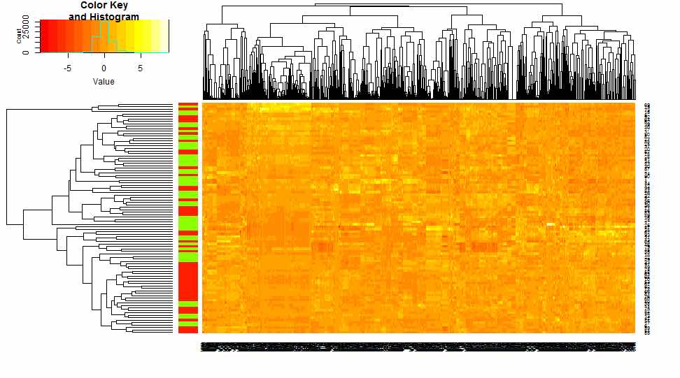
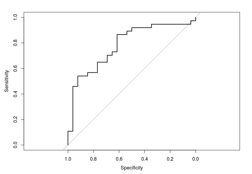
Total 871 imaging features of texture, wavelet and gray level histogram were extracted from the segment areas of OALs and IOIs in the primary cohort. Six features were selected according to the LASSO logistic regression. The features are the median of gray level, sum of average at 135°Gray Level Co-occurrence Matrix(GLCM), long run emphasis(LRE) of gray level run length matrix(GLRL), short run high gray emphasis (SRHGE) of GLRL, short run low gray emphasis(SRLGE) of GLRL and long run emphasis(LRE) of gray level run length matrix(GLRL). Five of six features were texture features (four from FS post-enhanced T1WI and one from T2WI) and one was wavelet feature (from T2WI). A good discriminating performance was shown based on combination of aforementioned six features with AUC of 0.81[95%CI, 0.77 to 0.84] in primary cohort and 0.78 [95%CI, 0.7 to 0.85] in validation cohort.Discussion
Traditional imaging differential diagnosis of OAL and IOI mainly relies on visual observation of radiologist, which is subjective and unstable. This research firstly analyses the radiomics MR imaging features of these two diseases which are objective and quantitive. The most useful discriminating features were provided and their predictive performance was proved to be good in validation cohort. These texture and wavelet features reflect different arrangement of components of different lesions. Radiomics features had shown promise in the prediction of treatment response, lymph node metastasis and assessing cancer genetics in previous studies [3, 4, 5]. Our research shows the value of imaging features in differentiating different diseases which resemble in conventional imaging characters. The limitation of current research is that more sequences and more imaging modalities should be involved.Conclusion
Radiomics imaging features of conventional multi-parametric MR protocol are conducive to a confirm diagnosis of OALs and IOIs. Both the texture and wavelet features based on T2WI and FS post-contrast T1WI contribute to the discrimination. The results are encouraging in showing the potential of imaging radiomics features in tissue identification and providing more information for treatment.Acknowledgements
This work was supported by the Beijing Municipal Administration of Hospitals Clinical Medicine Development of Special Funding Support (ZYLX201704); “Key Talent Project”of Beijing (2014001) ;“The priming scientific research foundation for the senior researcher in Beijing Tongren Hospital, Capital Medical University(TRYY-KYJJ-2016-011 ) .References
1 Woolf D K, Ahmed M, Plowman P N. Primary lymphoma of the ocular adnexa (orbital lymphoma) and primary intraocular lymphoma[J]. Clinical Oncology, 2012, 24(5): 339-344.
2 Wu A, Andrew N H, McNab A A, et al. IgG4-related ophthalmic disease: pooling of published cases and literature review[J]. Current Allergy and Asthma Reports, 2015, 15(6): 1-8.
3 Parmar C, Leijenaar RT, Grossmann P, Rios Velazquez E, Bussink J, Rietveld D, et al. Radiomic feature clusters and prognostic signatures specific for Lung and Head & Neck cancer. Scientific Reports. 2015;June 5(5):11044
4. YQ Huang,CH Liang,L He, et al. Development and validation of a Radiomics Nomogram for Preoperative Prediction of Lymph Node Metastasis in Colorectal Cancer. J Clin Oncol, 2016
5. Gutman D A, Dunn Jr W D, Grossmann P, et al. Somatic mutations associated with MRI-derived volumetric features in glioblastoma[J]. Neuroradiology, 2015, 57(12): 1227-1237.
Figures