2888
The Uesfulness of Intravoxel Incoherent Motion Diffusion-weighted Imaging for Predicting the Prognosis of Patients with Acute Myeloid Leukemia1Second Hospital of Shanxi Medical Hospital, Taiyuan, People's Republic of China, 2Radiology, Second Hospital of Shanxi Medical University, Taiyuan, People's Republic of China
Synopsis
Intravoxel Incoherent Motion (IVIM) is capable of providing both diffusion and perfusion quantification using a single imaging study at the same time, without the need for intravenous contrast injection. acute myeloid leukemia (AML) patients with complete remission and non-complete remission could exhibit different characterizations before treatment in perfusion and tissue cellularity of the lumbar bone marrow. This study was to investigate the usefulness of IVIM parameters in evaluation of prognosis in AML patients.
Author information
Jin-Liang Niu, Hong-Wei Wang, Xiao-Li Song Second Hospital of Shanxi Medical University, Taiyuan, ChinaPurpose
To investigate the value of Intravoxel Incoherent Motion (IVIM) parameters in evaluation of prognosis in patients with acute myeloid leukemia (AML) before treatment.Methods
Fifty-three patients before standard chemotherapy underwent MRI scans at 1.5T using conventional diffusion weighted imaging (DWI) and IVIM (b= 0,10,25,50,100,200,400,600,800,1000, 1200s/mm2) on sagittal planes covering the lumbar bone marrow. The IVIM parameters (perfusion fraction [f], molecular diffusion coefficient [D], and perfusion-related D [D*]) and apparent diffusion coefficient (ADC) were extracted on bone marrow. The percentage of leukemia cells in bone marrow was recorded, all patients was divided into complete remission (CR) and non-remission (NR) group.Results
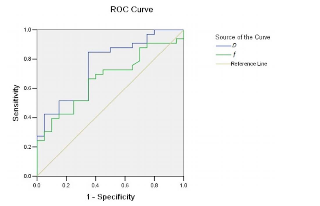
33 patients achieved CR and 20 patients NR based on the criteria for treatment response. The ADC values were not significant different between the two groups (p=0.378). However, D value of CR group was significantly higher (p=0.010), f value of CR group was significantly lower (p=0.021) than those of NR group. D* values had no significant differences between the two groups. The age of CR group was significantly higher than that of NR group (p=0.026). D value was negatively correlated with age. Using receiver operator characteristic (ROC) analysis, the area under the curve (AUC) of D and f were 0.759 and 0.666 respectively in evaluating prognosis of patients with AML before treatment.Conclusion
There were significant differences in IVIM parameters between CR and NR patients of AML before treatment, and the D and f were important values in evaluating prognosis of patients with AML.Acknowledgements
Thank for the technical support from Dandan Zheng, GE Healthcare MR Research, Beijing, China.References
[1]EsteyE,DöhnerH .Acute myeloid leukaemia. Lancet 2006 ; 368 ( 9550 ): 1894 – 1907 .
[2] Ferrara F ,Palmieri S , Leoni F . Clinically useful prognostic factors in acute myeloid leukemia .Crit Rev OncolHematol2008 ;66 ( 3 ): 181 – 193 .
[3]BourillonC, Rahmouni A, Lin C, et al.Intravoxelincoherent motion diffusion-weighted Imaging of multiple myeloma lesions: correlation with whole-body dynamic contrast agent-enhanced MR imaging[J].Radiology,2015, 277(3): 773-783.
[4]Koh DM, Collins DJ, Orton MR. Intravoxel incoherent motion in body diffusion-weighted MRI: reality and challenges. American Journal of Roentgenology ,2011,196(6):1351-1361.
[5]. Takahara T, Kwee TC. Low b-value diffusion-weighted imaging: emerging applications in the body. J MagnReson Imaging. 2012,35(6):1266-1273.
Figures
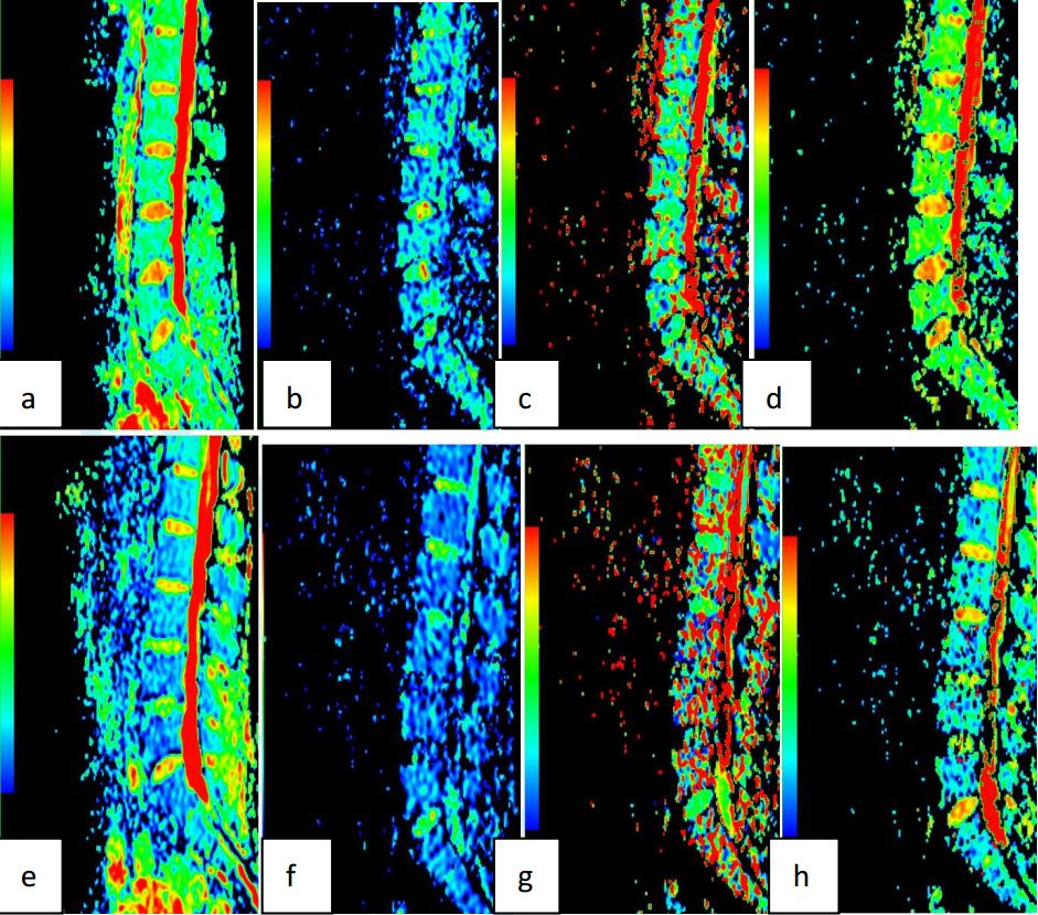
Figure1:(a-d) One patient with AML,male,25 Y, achieved CR after the first remission induction chemotherapy, (a) ADCmap, ADC=0.490×10⁻³mm2/s; (b) D map, D=0.243×10⁻³mm2 /s; (c) D*map, D*=66.0×10⁻³mm2 /s; (d) f map, f=0.201. (e-h) Another patient with AML, male,48 Y, achieved NR after the first remission induction chemotherapy:(e) ADCmap, ADC=0.468×10⁻³mm2 /s; (f) D map, D=0.199×10⁻³mm2 /s; (g) D*map, D*=64.8×10⁻³mm2 /s;(h) f map, f=0.281. Color bars of the two patients are the same.