2869
One Minute Free-Breathing 3D Cardiac Cine Imaging with Adaptive Respiratory Self-Gating Efficiencies1University of California San Francisco, San Francisco, CA, United States, 2New York University, New York, NY, United States
Synopsis
Cardiac cine imaging has become the standard for cardiac functional measurements. However, a series of breath-holds are required to acquire 2D cine images covering the whole heart. The capability of children or sick patients to perform consistent breath-holds is limited and often results in non-diagnostic images. We aim to develop a fast and reliable 3D imaging technique for cardiac functional assessment, which only requires one minute of scan time during free breathing. To compensate for respiratory motion, which varies substantially among subjects, we propose to apply adaptive respiratory self-gating efficiencies to generate reliable image quality for 3D cardiac cine imaging.
INTRODUCTION
Free breathing 3D cardiac
cine imaging techniques have been developed to overcome limitations of the
current standard breath-hold 2D cine imaging. However, the associated scan time
increases significantly with improved imaging settings and image quality relies
heavily on compensation for respiratory motion, which can vary substantially
between different patients. A robust and reliable method is highly desirable.
In this study, we propose to achieve 3D cine imaging in one minute during free breathing,
with improved motion compensation by using adaptive respiratory self-gating
efficiencies. MATERIALS AND METHODS
Previously we have developed
an accelerated free-breathing 3D cardiac cine imaging technique [1] using a
pseudo-random variable-density undersampling strategy, called CIRcular
Cartesian UnderSampling (CIRCUS) [2-4]. Our previous results have demonstrated
that choosing respiratory gating efficiency between 25%-50% can generate
reasonable motion compensation for a scan of 2.5-3 mins using the CIRCUS
acquisition strategy and an image reconstruction method that combines
compressed sensing and parallel imaging (the so called k-t SPARSE-SENSE method)
[5,6]. In this study, we propose to optimize the motion compensation strategy
while further reducing scan time.
Free breathing 3D cine imaging covering the entire
left ventricle in a short axis view was acquired on 14 healthy volunteers on a
3.0T MR scanner (GE Medical Systems, Milwaukee, WI) with an 8-channel cardiac
coil. The imaging parameters were: FOV = 34.0×25.5 cm2, TR/TE =
4.1/1.7ms, flip angle = 60°, readout bandwidth = ± 125kHz, slice thickness = 4-5
mm, image matrix = 256×144, and number of slices = 28-30. The scan time for 3D
imaging was of the order of 2.5 mins. ECG triggers detected during the 3D scan were
saved for retrospective gating in the image reconstruction. Temporal resolution
was chosen to be 10×TR=41ms.
With the variable-density CIRCUS acquisition, the
k-space central line along kx
(ky=kz=0) was repeatedly acquired and cross-correlation of
corresponding signals was performed to obtain a respiratory motion signal. The
principle correlation of that data was derived by applying principle component
analysis (PCA), which yielded a 1D signal (self-gating signal) containing motion
information within the FOV. The derived motion signal was used for sorting the
3D data into a set of bins according to the displacement of the self-gating
signal. 3D cine images with the acquired data in the entire 2.5 mins scan time
were reconstructed as the reference, with a fixed respiratory gating efficiency
of 25%.
We retrospectively mimic a shorter scan time of 1 min,
by only using the data during the first 1 minute of the scan. Instead of
choosing a fixed gating efficiency for the 1 min data sets, we used a series of
gating efficiencies by accumulating the bins one by one (so called “adaptive”).
All the data sets with different efficiencies were included in the image
reconstruction. Thus, instead of discarding some data as happens in
conventional gating, all the data was included in the reconstruction, which not
only provides adaptive gating efficiencies but also improves the data fidelity.
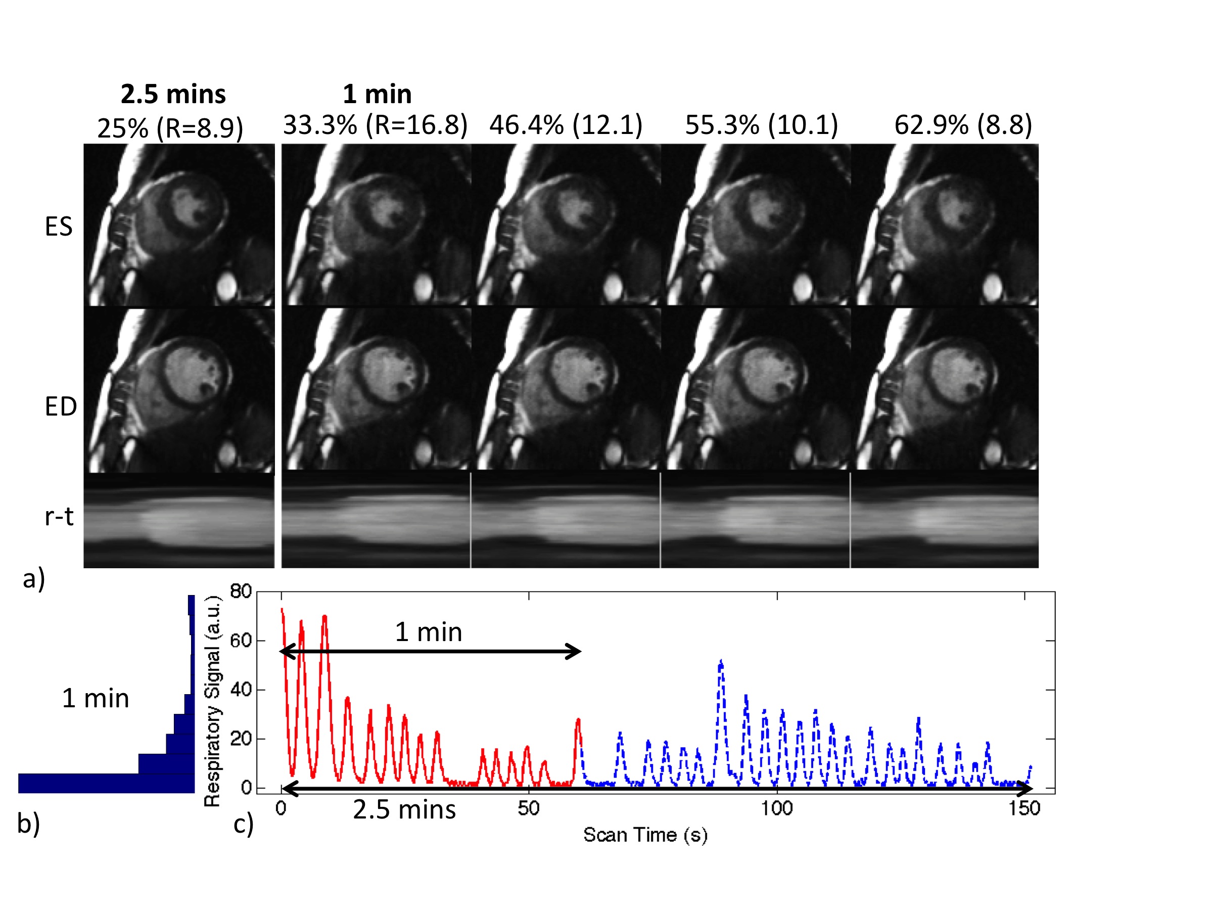
RESULTS AND DISCUSSION
We selected data for review
from 3 subjects whose heart rates were similar (~70 bpm) but with different
respiratory motion. With the derived self-gating signal, data was sorted into
bins and the data distribution varied according to the individual breathing
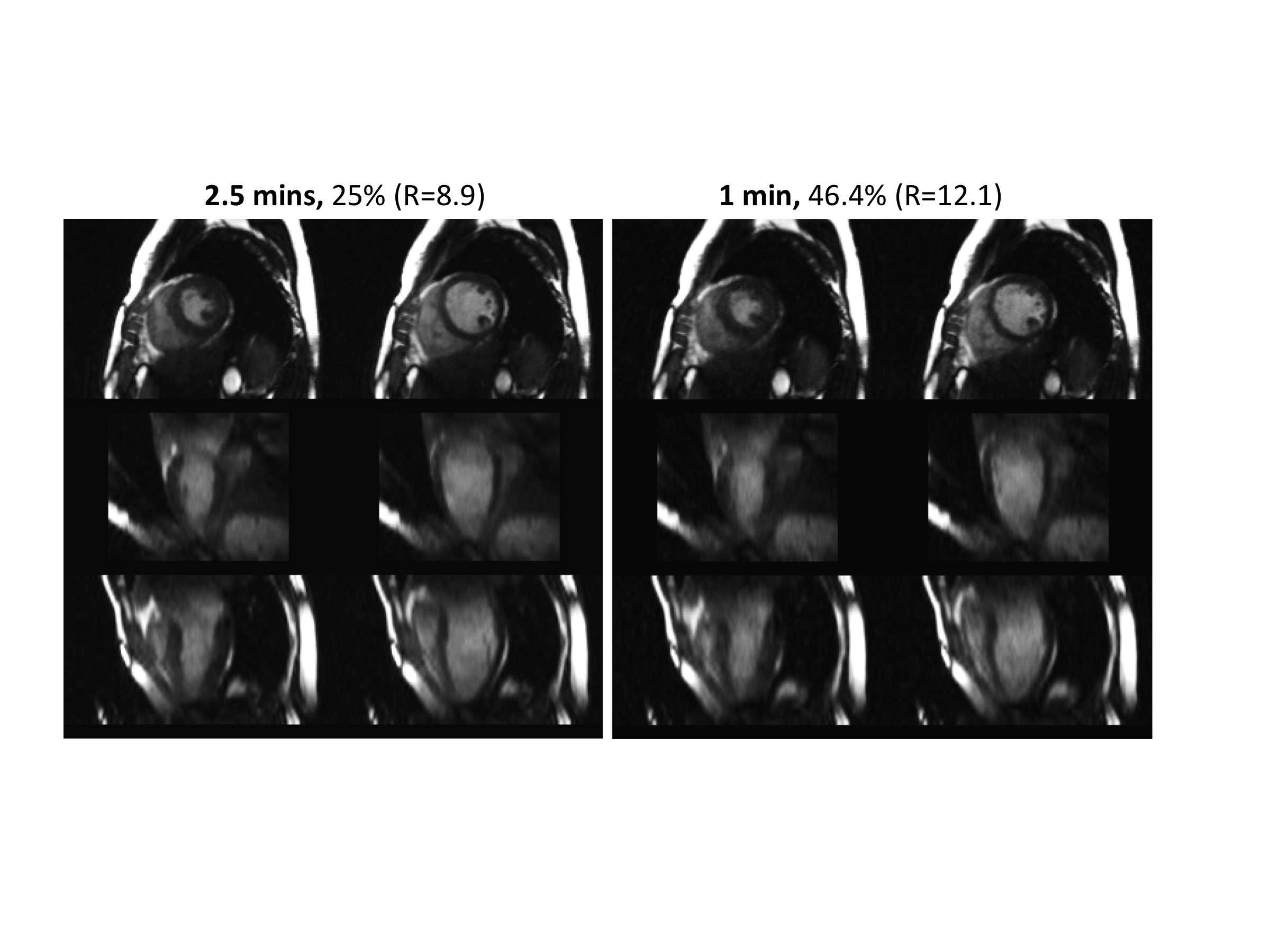
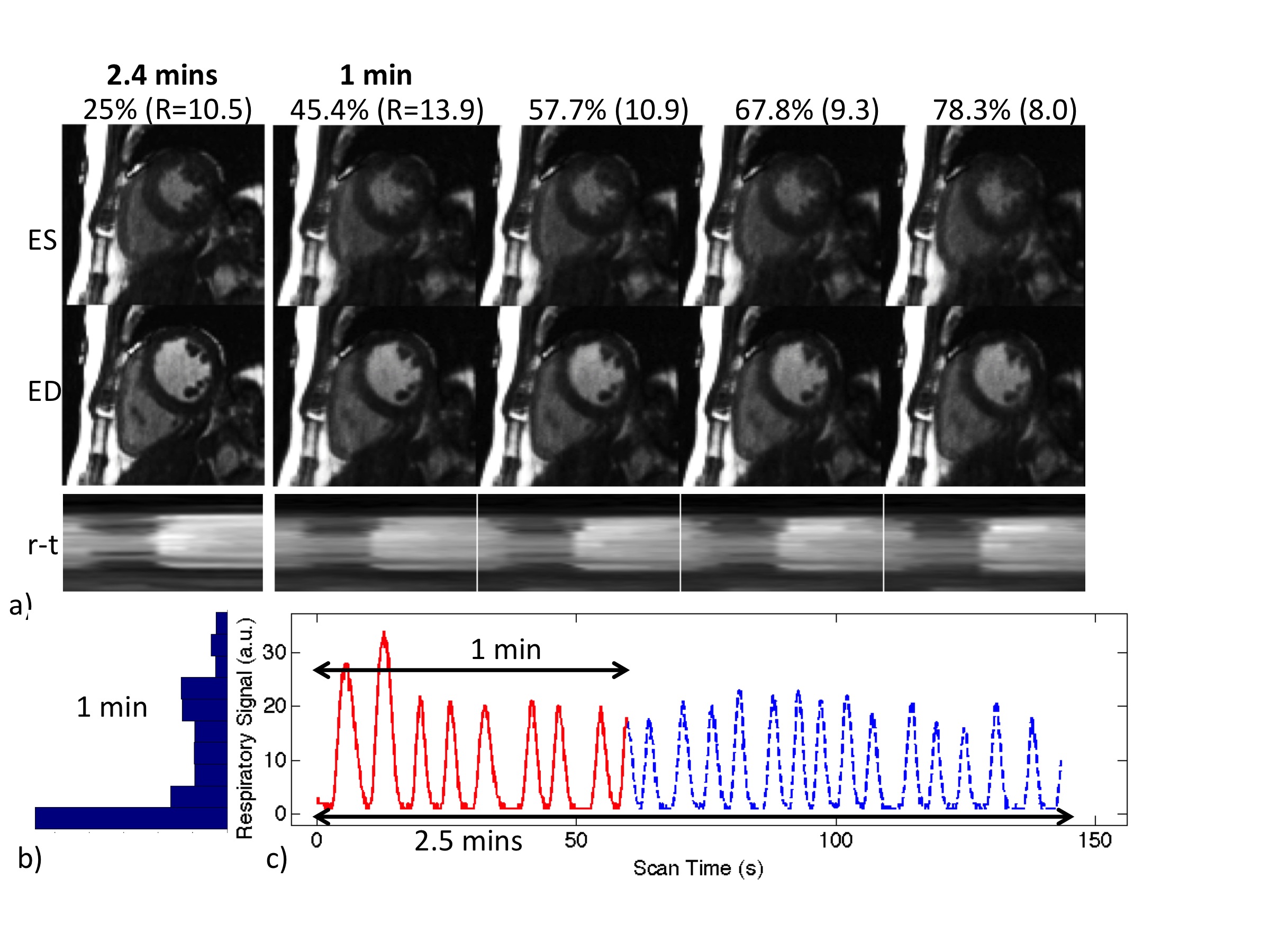
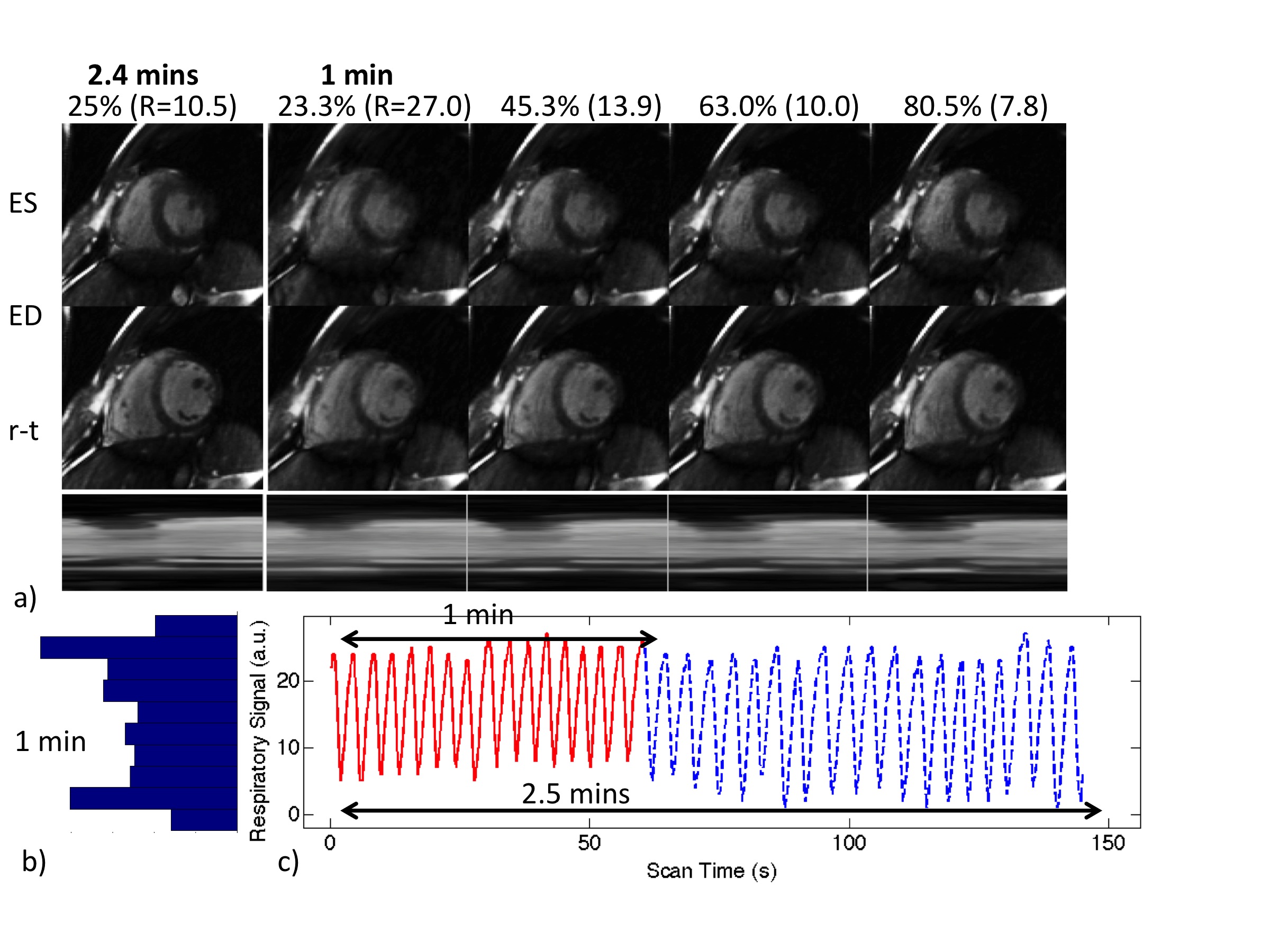
patterns, which directly affects the image quality. Three cases are shown here
(Figs. 1-4), with images from 2.5 mins and 1 min scans, at end-systolic (ES) and
end-diastolic (ED) phases, and the signal
profile across the left ventricle throughout the cardiac cycle (r-t). With the
proposed adaptive respiratory gating efficiencies, we generated high
quality and reliable 3D cardiac cine images even for data from one minute of
acquisition, regardless of the varying respiratory motion patterns. CONCLUSIONS
We have developed a highly
accelerated, free breathing, 3D cardiac cine imaging approach which could be completed
in one minute using adaptive respiratory self-gating efficiencies. This method
can provide a significantly improved MR imaging tool for assessing cardiac function. Acknowledgements
NIH K25 EB014914 (JL), NIH R56HL133663 (JL), GE Healthcare Research Grant (JL)References
1. Liu J, Feng L, Saloner D. Highly Accelerated Free-breathing 4D Cardiac Imaging with CIRCUS Acquisition. The 22nd Annual Meeting of ISMRM 2014, Milan, p429.
2. Liu J, Saloner D. Accelerated MRI with CIRcular Cartesian UnderSampling (CIRCUS): a variable density Cartesian sampling strategy for compressed sensing and parallel imaging. Quant Imaging Med Surg. 2014 Feb; 4(1):57-67.
3. Liu J, Pedoia V, Heilmeier U, Ku E, Su F, Khanna S, Imboden J, Graf J, Link T, Li X. High-temporospatial-resolution dynamic contrast-enhanced (DCE) wrist MRI with variable-density pseudo-random circular Cartesian undersampling (CIRCUS) acquisition: evaluation of perfusion in rheumatoid arthritis patients. NMR Biomed. 2016 Jan; 29(1):15-23. PMID: 26608949.
4. Liu J, Koskas L, Faraji F, Kao E, Wang Y, Haraldsson H, Kefayati S, Ahn Sinyeob, Laub G, Saloner D. Highly Accelerated 4D Flow for Intracranial Aneurysm Imaging. ISMRM Workshop on Quantitative MR Flow, San Francisco, Oct 2016.
5. Otazo R, Kim D, Axel L, Sodickson DK. Combination of compressed sensing and parallel imaging for highly accelerated first-pass cardiac perfusion MRI. Magn Reson Med. 2010;64(3):767-76. Epub 2010/06/11. doi: 10.1002/mrm.22463. PubMed PMID: 20535813; PubMed Central PMCID: PMC2932824.
6. Feng L, Srichai MB, Lim RP, Harrison A, King W, Adluru G, Dibella EV, Sodickson DK, Otazo R, Kim D. Highly accelerated real-time cardiac cine MRI using k-t SPARSE-SENSE. Magn Reson Med. 2013;70(1):64-74. doi: 10.1002/mrm.24440. PubMed PMID: 22887290; PubMed Central PMCID: PMC3504620.
Figures