2848
Evaluation of three-dimensional flow characteristics behind the prosthetic mechanical valve under subvalvular pannus formation; in-vitro 4D flow MRIHyung Kyu Huh1, Hojin Ha1, Sang Joon Lee1, Namkug Kim2,3, and Donghyun Yang3
1Mechanical Engineering, POSTECH, Pohang, Korea, Republic of, 2Department of Convergence Medicine, Asan Medical Center, 3Department of Radiology, Asan Medical Center
Synopsis
A pannus formation is a growth of abnormal tissues around the heart valve, which is often found in patients whom underwent heart valve replacement surgery. Significant hemodynamic changes caused by the formation of the pannus may contribute to the failure of the valves even without direct contact to the leaflets. In current study, flow in the sinus if Valsalva under subvalvular pannus formation was analyzed using in vitro phantom and 4D flow MRI. Symmetric pannus causes imperfect opening the valve, which might lead to the heart failure.
Introduction
A pannus formation is a growth of abnormal tissues around the heart valve, which is often found in patients whom underwent heart valve replacement surgery. Formation of pannus may dysfunction the replaced valves, which impose repeated open surgery of the heart[1]. Subprosthetic panni formation in patients with chest discomfort after valve replacement was observed by Han et al.[2] using cardiac computed tomography. Apart from the direct contact of pannus with prosthetic valves, significant hemodynamic changes may also contribute to the failure of the valves even without direct contact to the leaflets. However, due to the limitation in in vivo visualizing of the flow characteristics, dysfunction related with the flow characteristics are not fully understood yet. In current study, flow in the sinus if Valsalva under subvalvular pannus formation was analyzed using in vitro phantom and 4D flow MRI.Methods
An in vitro sinus phantom which geometry was based on the human data used in the study of Yagi et al.[3] was printed using 3D printer(Objet500 connex3, Stratasys, Eden Prairie, MN, USA) (Fig.1). The St-Jude prosthetic valve of 19.6mm diameter was placed in front of the aortic sinus. Two different types of pannus (symmetric and asymmetric) was installed 5mm in front of the prosthetic valve. T/R=0, 0.1, 0.2, 0.3, 0.4 and 0.5, where T is thickness of the pannus and R is the radius of the tube was used in current experiment. Steady flow rate of 6 and 12 L/min was applied, which represents mean flow rate and systolic flow rate in human aorta, respectively. Pulsatile flow was also applied in 60bpm. Image acquisition was performed on 3T scanner (MAGNETOM Skyra, Simens, Germany) with velocity encoding range (VENC) of 80 and 180 for 6 and 12 L/min, respectively.Results
Three distinctive jet flow of which velocity was higher than 41cm/s was found behind the prosthetic mechanical valve without pannus formation (T/R=0). Valve was widely opened, and recirculating flow was observed inside apices (Fig.2a). When symmetric pannus with T/R=0.5 was placed in front of the prosthetic valve, jet flow was developed toward the upper and lower parts of the Sinus and strong regurgitating flow was found in the center region of the Sinus (Fig.2b). Regurgitating flow was also confirmed at the pulsatile flow condition, indicating the failure of the valve function (data not shown). Lower jet was significant than upper jet for the case of asymmetric pannus with T/R=0.5 as shown in Fig.4a. As the asymmetric pannus was rotated for 45 degree, jet is shifted toward the side wall where pannus was not formed (Fig.4b). The shifting of the jet was more significant as the asymmetric pannus was rotated 90 degree.Discussion
Regardless of direct contact of pannus with the leaflets, failure of the prosthetic mechanical valve occurs when symmetric pannus is formed. This will increase the pressure inside the heart and it may cause heart failure. Also, high wall shear stress applied on the upper and lower walls of the Sinus may cause dilation. Imperfect opening of the valve is mainly due to the center focused flow formed behind the pannus. Leaflets of the bileaflet valves are fixed with the hinges, and it is opened when momentum is applied on the cross section area near the peripheral. However, center focused flow transfer its momentum on the central region of the valve, which compensates the opening momentum to maintain valves to be partially opened. Also, large recirculating flow in the center of the sinus applies resistant force to the valve opening. For the case of asymmetric pannus, strong recirculation at the upper apex press down the upper leaflet, which leads to the opening of the valve. Also, formation of the rotated asymmetric pannus applies non-axisymmetric wall shear stress, which can cause asymmetric Sinus dilation.Conclusion
Pannus formation and its shape, severity and rotational angle have great influence on the flow characteristics inside the Sinus. High wall shear stress applied on the wall may lead the dilation of Sinus. More importantly, flow characteristics plays significant role on the function of the prosthetic valve, which may cause heart failure. 4D MRI have strong potential to visualize and quantify 3D flow inside the sinus of real patient with prosthetic heart valve. Understanding the flow characteristics will be helpful to decide whether patient requires repeated open surgery.Acknowledgements
No acknowledgement found.References
[1] Chung MS, Yang DH, Kim DH, et al. Eur Heart J-Card Img (2015) [2] Han K, Yang DH, Shin SY, et al. Radiology (2015) [3] Yagi T, Yang W, Umezu M. Journal of Biomechanical Science and Engineering (2011)Figures
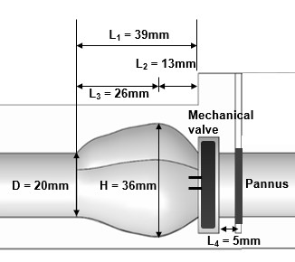
Sinus of Valsalva model and dimensions.
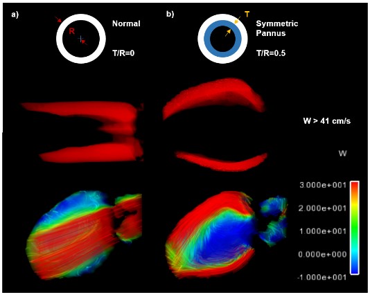
Jets
and streamline of symmetric pannus with a) T/R=0 and b) T/R=0.5.
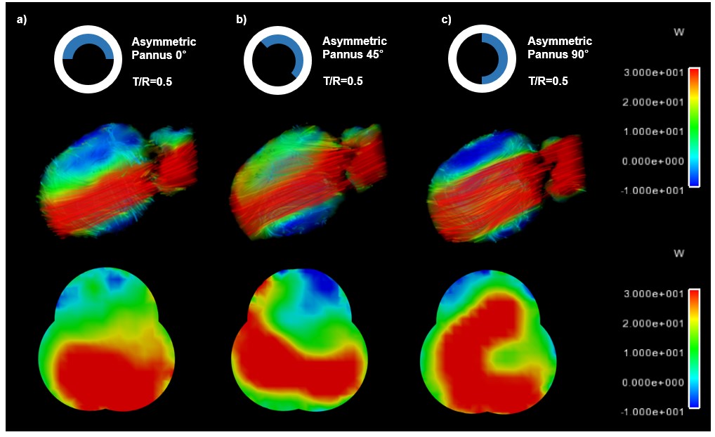
Sagittal
and axial view of velocity of asymmetric pannus (T/R=0.5) with rotation angle
a) 0, b) 45 and C) 90.