2847
Feasibility of non-contrast-enhanced cardiovascular 4D flow MRI using a balanced SSFP approach1Department of Electrical Engineering, Stanford University, Stanford, CA, United States, 2Department of Radiology, Stanford University, CA, United States
Synopsis
Balanced steady state free precession (bSSFP) phase-contrast sequences have desirable tissue contrast properties that allow for non-contrast-enhanced cardiovascular flow exams. However, they are susceptible to flow-related signal dephasing especially in regions with highly accelerating flow like the heart. To address this, we propose a variable-density radial view-ordered bSSFP 4D flow sequence. Acquired images show reasonable visualization of cardiac anatomy, and similar velocity measurements in aortic regions. Further sequence modifications are suggested to improve its robustness. If proven to be a viable alternative to the standard exam, bSSFP 4D flow would reduce exam costs and greatly improve patient experience.
Purpose
Volumetric, time-resolved phase-contrast (4D flow) MRI data is traditionally acquired with an RF-spoiled gradient echo (SPGR) sequence which produces T1-weighted tissue contrast. For this reason, T1-shortening intravenous contrast agents such as gadolinium or ferumoxytol are required to simultaneously produce strong blood signal, assess anatomy, and precisely measure flow. However, use of contrast agents increase exam costs and reduce patient comfort especially for pediatric cases. Balanced steady state free precession (bSSFP) phase-contrast sequences sidestep the need for contrast agents by providing T2/T1 tissue contrast1-4, but require extra care in maintaining the steady state and mitigating flow-related signal dephasing5. Nielsen and Santini have previously proposed bSSFP 4D flow sequences for slower flow applications in the head and neck. In this work, we implement a radial view-ordered bSSFP 4D flow sequence, and show its feasibility in non-contrast-enhanced flow imaging of the heart.Methods
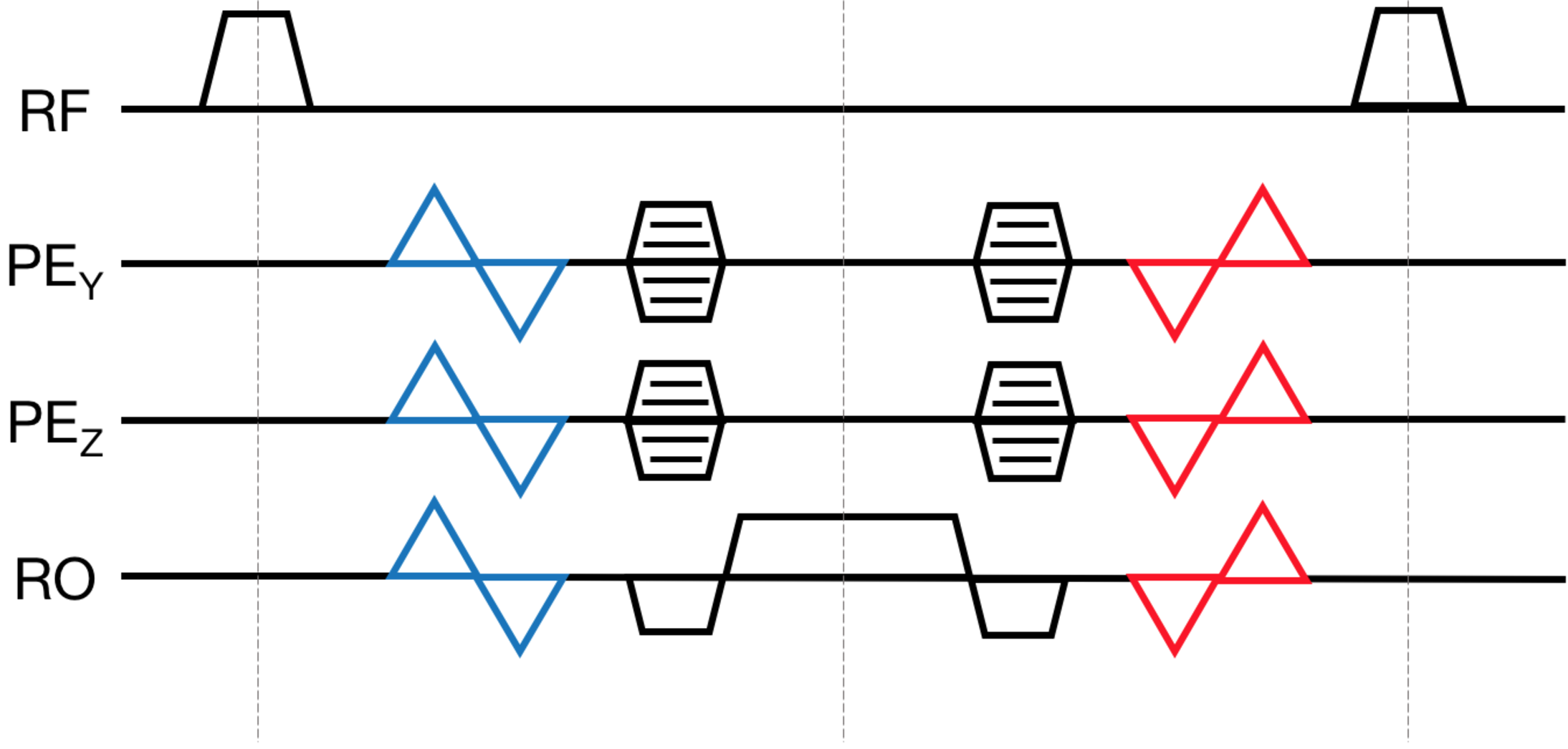
The proposed sequence diagram is shown in Figure 1. Velocity encoding is done using a simple 4-point scheme with interleaved flow encoding. A non-selective hard-pulse excitation and combined bipolar/phase encode gradients are utilized to make TE and TR as short as possible. The volumetric Cartesian dataset is acquired using variable-density center-out radial view ordering6 to keep phase encode amplitudes small, and consequently reduce first moment contributions that could disrupt the steady state.
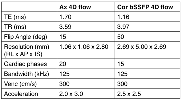
Scanner Experiments: All experiments were performed on a 3T General Electric MR 750 using a 32-channel cardiac coil. Static phantoms with varying concentrations of gadobutrol were scanned to evaluate the contrast properties of the bSSFP 4D flow. With IRB approval and informed consent, a pediatric patient referred for a gadolinium-enhanced cardiac 4D flow MRI was recruited for this study. A non-contrast-enhanced bSSFP 4D flow was performed followed by a standard post-contrast SPGR 4D flow. Scan parameters for both acquisitions are described in Figure 2. A parallel imaging and compressed sensing based scheme was implemented in BART and used to reconstruct both datasets7.
Results
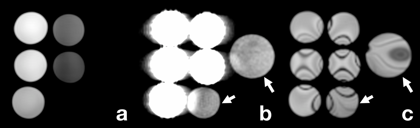
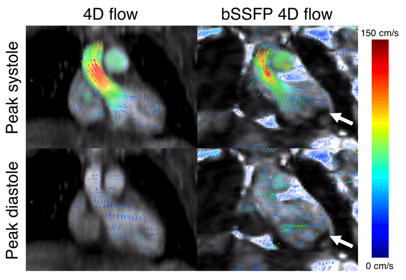
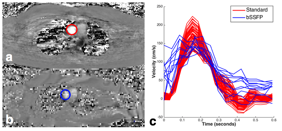
As shown in Figure 3, the static phantom showed the desired contrast properties of a balanced SSFP sequence. Despite the lack of contrast administration, the in-vivo bSSFP images shown in Figure 4 produced good visualization of cardiac anatomy, and reasonably similar velocity measurements in the aortic valve when compared to the standard 4D flow experiment. However, velocities are not able to be resolved in the right and left apical ventricular blood pools due to the presence of banding artifacts.Discussion
These preliminary results indicate that cardiovascular bSSFP 4D flow is feasible, although signal loss and image artifacts due to steady-state disruptions may limit its robustness. There is a reduction in precision and accuracy of measured velocities in phase encode directions (Figure 5), which can be attributed to steady state disruptions possibly caused by non-zero first gradient moments. For complete flow-compensation over TR, higher moment bipolar gradients must be played after readout; although, they will drag out the TR and introduce more banding artifacts. We believe these can be mitigated by scanning at lower field strengths, higher-order shimming8, and/or phase-cycled acquisitions9. Further signal variations are introduced by TR-to-TR variations in flow and phase encoding gradients, but can be mitigated using a reverse centric radial view-ordering and non-interleaved flow encodes. Finally, adding extra flow-compensating bipolar gradients increases second gradient moments by a factor of ~10 (depending on Venc and FOV), which could lead to further steady state disruption in the presence of highly accelerating flow. Further investigation is necessary to fully characterize second moment effects.Conclusion
Our results show that a non-contrast-enhanced balanced SSFP 4D flow exam may eventually be a viable alternative to the standard 4D flow. Such a sequence would obviate the need for intravenous contrast agents, reducing exam costs, and improving patient experience.Acknowledgements
National Science Foundation Graduate Research Fellowship (DGE-114747), the Tashia and John Morgridge Faculty Scholars Fund, and GE HealthcareReferences
[1] WR Overall, DG Nishimura, BS Hu. Fast phase-contrast velocity measurement in the steady state. Magn Reson Med, 2002; 48:890-98.
[2] M Markl, MT Alley, NJ Pelc. Balanced phase-contrast steady-state free precession (PC-SSFP): A novel technique for velocity encoding by gradient inversion. Magn Reson Med, 2003; 49(5):945-52.
[3] JF Nielsen, KS Nayak. Referenceless phase velocity mapping using balanced SSFP. Magn Reson Med, 2009; 61(5):1096-102.
[4] F Santini et al. Time-resolved three-dimensional (3D) phase-contrast (PC) balanced steady-state free precession (bSSFP). Magn Reson Med, 2009; 62(4):966-74.
[5] O Bieri & K Scheffler. Flow compensation in balanced SSFP sequences. Magn Reson Med, 2005; 54:901-07.
[6] JY Cheng et al. Variable-density radial view-ordering and sampling for time-optimized 3D Cartesian imaging. In Proceedings of the ISMRM Workshop on Data Sampling and Image Reconstruction, Sedona, Arizona, USA, 2013.
[7] M Uecker, F Ong, JI Tamir et al. Berkeley Advanced Reconstruction Toolbox. In Proceedings of the 23rd Annual Meeting of ISMRM, Toronto, Ontario, Canada, 2015.
[8] DH Kim et al. Regularized higher-order in vivo shimming. Magn Reson Med, 2002; 48(4):715-22.
[9] A Datta, et al. Breath-held phase-cycled cardiac CINE MRI using slow frequency modulation. Proceedings of the 24th Annual Meeting of ISMRM, Singapore, 2016.
Figures