2845
Improving Non-Contrast 4D Flow MRI Using Multiple-Thin-Slab Dual-Venc Acquisition1Technical University of Munich, Munich, Germany, 2GE Healthcare, CA, United States, 3GE Global Research, Munich, Germany, 4MRI Research Center, University Children Hospital, Zurich, Switzerland
Synopsis
Dual-venc 4D flow MRI can improve velocity-to-noise ratio (VNR) using a low-venc velocity map to provide a good VNR and a high-venc velocity map to correct the velocity aliasing. However, dual-venc 4D flow MRI significantly lengthens the scan duration to longer than the 10min delayed enhancement wait time and therefore it is difficult to take advantage of T1 shortening immediately after contrast injection. On the other hand, whole volume acquisition saturates blood signal and decreases SNR for 4D flow MRI, especially without contrast agent. Multi Thin Slab (MSLAB) 4D flow MRI acquisition technique provides bright blood imaging with improved SNR and motion robustness. Therefore, by combining dual-venc with MSLAB we propose a more accurate 4D flow method without requirement of contrast agent. In this study, we analyzed our proposed method in a pulsatile flow phantom in terms of VNR and performance in a volunteer.
Purpose
4D flow enables comprehensive assessment of hemodynamics-related cardiovascular pathologies in the entire chest. However, its accuracy is limited without administration of T1-shortening contrast agents due to the low SNR in blood pool. This work aimed to improve non-contrast 4D flow through the combination of two methods: increasing velocity-to-noise ratio (VNR) by dual-venc encoding1,2, and increasing SNR by applying Multi Thin Slab (MSLAB) acquisition3.Methods
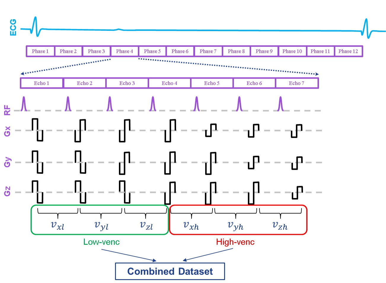
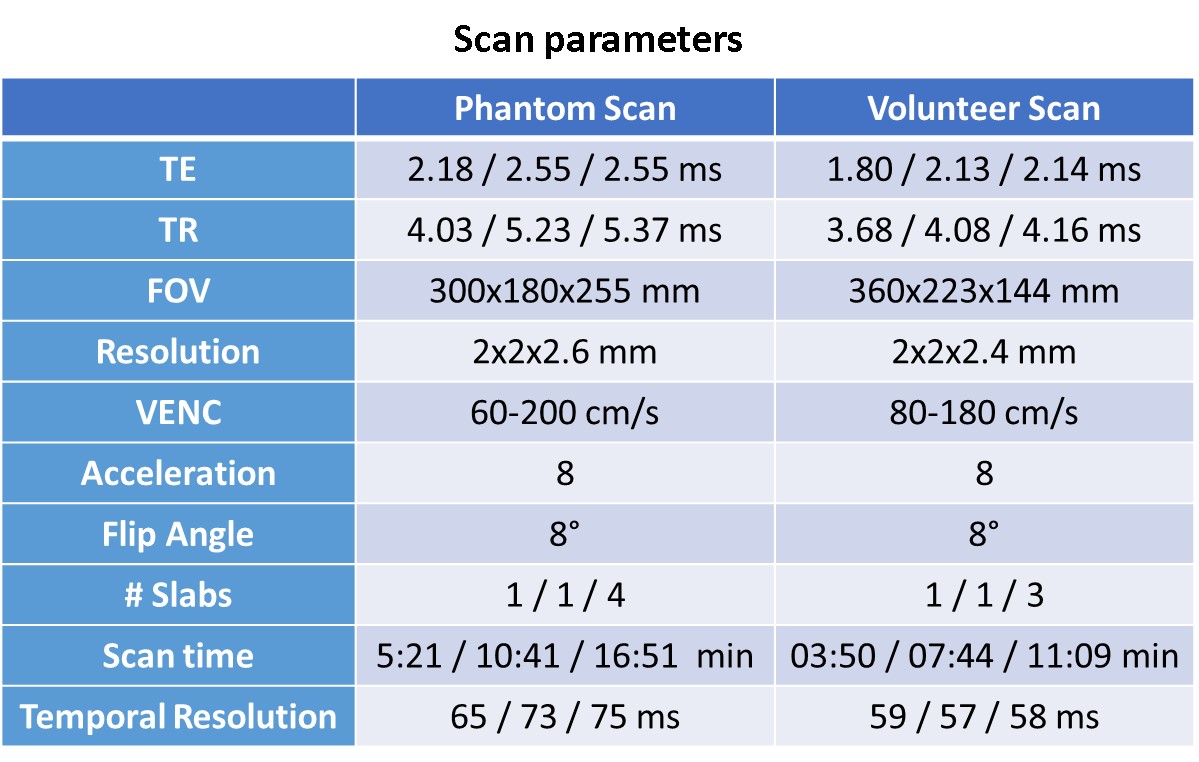
The acquisition used Cartesian variable density random k-t sampling3 with imaging parameters shown in Table 1 and image was reconstructed using kat-ARC4. For MSLAB, different slabs were reconstructed separately and then volume-merged using weighted averaging on the overlapping slices according to their slab-center distances.3 Dual-venc 4D flow MRI was implemented as shown in Fig.1. Maxwell term and eddy current correction was performed in post-processing for each velocity image. The high VNR velocity image was obtained from the low-venc image, with phase wrapping corrected based on the high venc image exempt from velocity aliasing.
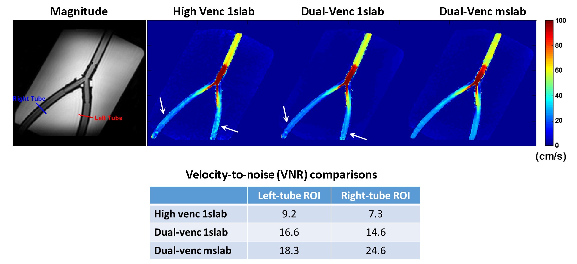
Experiments: The closed-circuit pulsatile flow phantom was driven by an industrial membrane flow pump with heart rate 61 beats/min and scanned on a GE 3T MR750w MR scanner (Waukesha, WI, USA). As shown in Fig 2, the phantom contains a Y-shaped tube filled with non-contrast blood mimicking fluid (T1=1600ms) and was placed in an oblique position.5 Then, VNR was calculated at an ROI using $$$VNR= (v_1 + v_2)⁄(√2 std(v_1-v_2))$$$, where $$$v_1$$$ and $$$v_2$$$ are the velocities in identical ROIs from the repetitive measurements1. As shown in Fig.2, VNR was evaluated at two ROI’s near tube outlet. A volunteer was scanned without contrast agent on GE 3T MR750 (Waukesha, WI) for whole-chest 4D flow MRI. The images were reconstructed on MATLAB (MathWorks) and processed on Arterys (San Francisco, CA, USA).
Results
Fig. 2 shows the velocity images of 1SLAB single-venc, 1SLAB dual-venc and MSLAB dual-venc 4D flow MRI from flow phantom experiments and comparisons of VNRs in the selected ROI’s. Dual-venc 4D flow MRI improves VNR and shows less noisy flow field, especially in the low-velocity regions. MSLAB dual-venc 4D flow MRI provides higher signal in the flow tissue due to inflow enhancement and thus further increases VNR compared to 1SLAB dual-venc.
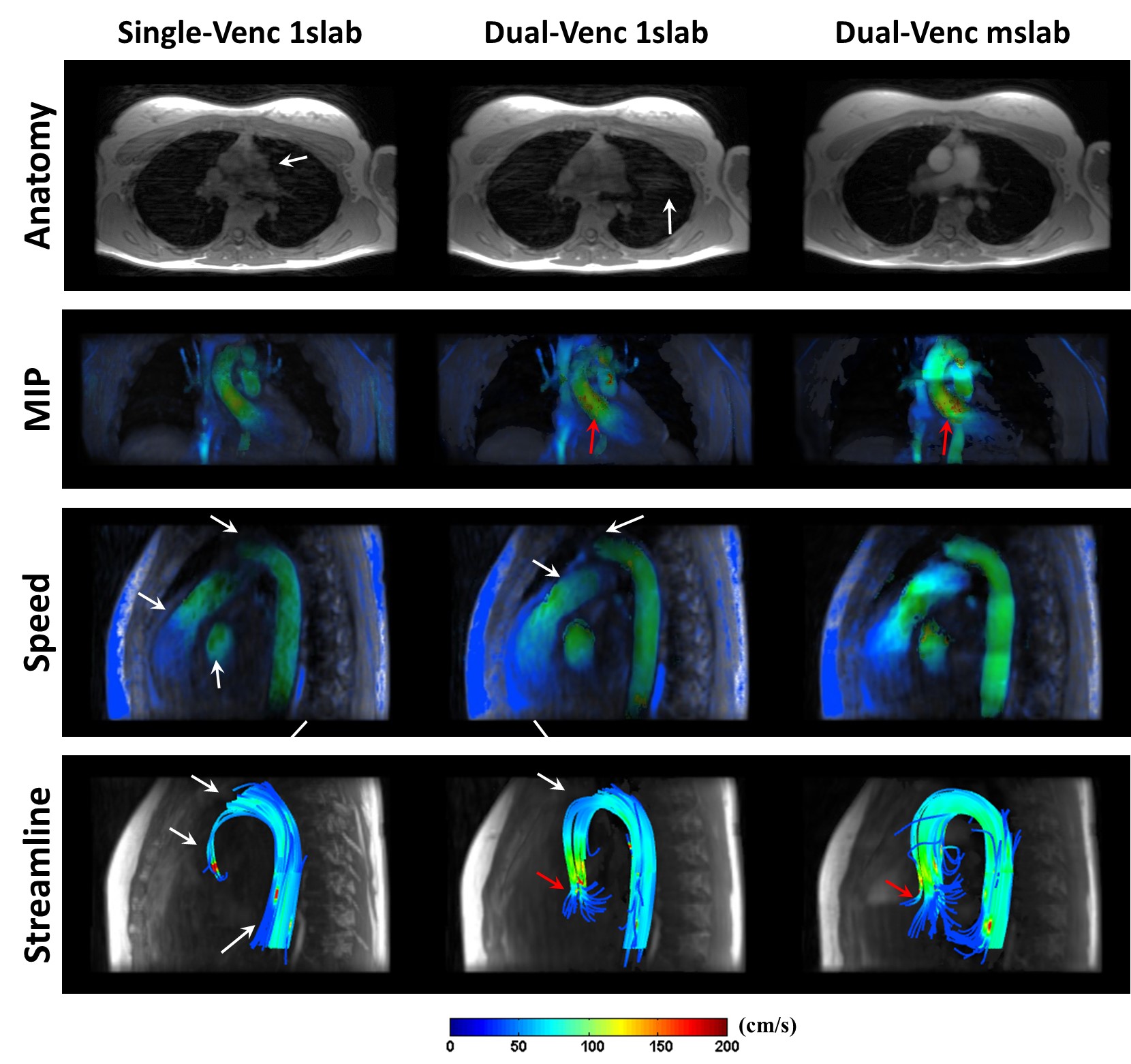
Similarly, dual-venc 4D flow MRI improves VNR on the volunteer especially in low velocity regions such as right and left pulmonary arteries and ventricles as shown in Fig. 3. Moreover, MSLAB dual-venc 4D flow MRI improves velocity image quality for the acquisition without non-contrast agent due to the blood enhancement. These improvements are visible in the aortic arc, pulmonary arteries and left-ventricle (Fig.3). Also, with much brighter blood, the flow field obtain using MSLAB is less sensitive to residual respiratory motion artifacts than 1Slab.
Discussion & Conclusion
With dual-venc acquisition, higher first gradient moments are needed to achieve the low venc than single venc and it increases the gradient level and TE. The longer TE introduces more signal loss related to dephasing due to the turbulence or higher order motions. In Fig.3 (shown with red arrow), we can see the phase dispersion especially at aortic root near the valve because of turbulence. In future work, we plan to use high-venc dataset in the regions of turbulence.
For non-contrast scans, MSLAB 4D flow MRI provides bright-blood anatomical images without slab misregistration and with less visible motion artifacts.3 High contrast between blood and background tissues helps vessel segmentation needed for accurate flow measurements.
This study shows that velocity mapping with improved VNR is feasible for non-contrast whole-chest 4D flow with the proposed dual-venc multiple thin slab acquisition.
Acknowledgements
This work was funded by the European Commission under Grant Agreement Number 605162.References
1. Nett EJ et al., Four-Dimensional Phase Contrast MRI With Accelerated Dual Velocity Encoding, J Magn Reson Imaging. 2012 Jun;35(6):1462-71.
2. Susanne Schnell et al., ISMRM 2015: 4546
3. Lai P et al., ISMRM 2016: 2709
4. Lai P et al., ISMRM 2009:767
5. Hafalir et al., ISMRM Workshop on Quantitative MR Flow 2016
Figures