2842
Improved Visualization of Common Iliac Artery 3D-cine Phase Contrast Magnetic Resonance Imaging Using Selective Water Excitation1Department of Radiological Technology, Kurashiki Central Hospital, Okayama, Japan, 2Philips Electronics Japan, Tokyo, Japan, 3Department of Medical Technology, Kurashiki Central Hospital, Okayama, Japan
Synopsis
The motion artifacts in the pelvic area using 3D-cine phase contrast (4D PC) MRI may be provide the severity results. The purpose of this study was to demonstrate the usefulness of fat suppression for the common iliac artery (CIA). The study protocol compared 4D PC MRI date without fat suppression to with principle of selective excitation technique (ProSet). We evaluated the streamline visualization, SNR of magnitude images and blood flow volume. ProSet can reduce respiration artifacts. Extending of TR by using ProSet increases SNR. Thus ProSet1-1 is the optimal fat suppression technique to improve vascular visualization.
Purpose
Recently, magnetic resonance fluid dynamics (MRFD) using 4D-PC MRI has been reported as a method for obtaining hemodynamic information. Several studies have reported that 4D-PC MRI after contrast agent injection especially at 3T MR system significantly improved signal-to-noise ratio (SNR) in magnitude date and 3D flow visualization 1.2. In the thoracic aorta and pulmonary artery, navigator gating can be used to reduce artifacts and provide sensitive hemodynamic analysis 3. Commonly, since navigator or respiratory gating cannot be often used in the pelvic area, it suffers from severe motion artifacts. To reduce such motion artifacts in the pelvis area, we tried to combine fat suppression (selective water excitation) technique. The purpose of this study was to demonstrate the usefulness of fat suppression for the common iliac artery (CIA).Methods
Three healthy volunteers (age=33.3 ± 9.7) participated in this study. We obtained IRB approval and informed consent from all subjects. All imaging protocols were done at 1.5T scanner (Ingenia, Philips Healthcare, Best, The Netherlands). The 4D-PC data was acquired retrospectively with PPU-gating and without respiratory gating. Imaging parameters were: acquisition matrix, 2.08*2.08*4mm; reconstruction matrix, 1.37*1.37*4mm; flip angle, 10; No. of phase,12 ) To analyze the obtained 4D-flow data, we used blood flow analysis software (GTFlow, GyroTools, Zurich, Switzerland) First, we compared several images types (without water excitation, ProSet1-1 and ProSet1-2-1) for the image quality of the streamline visualization, and measured SNR of magnitude images. Subsequently, we compared the optimal water excitation method with conventional sequence to validate the effect of fat suppression. We compared the blood flow volume based on the abdominal aorta and CIA velocities obtained by both 2D of reference standard and 4D-PC MR with water excitation.Results
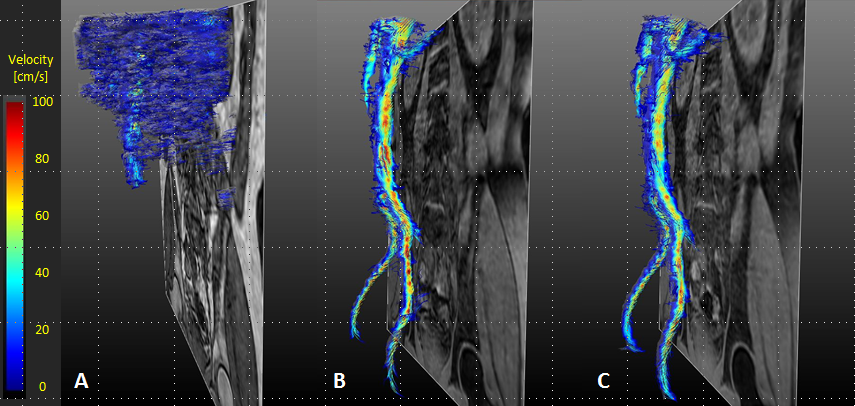
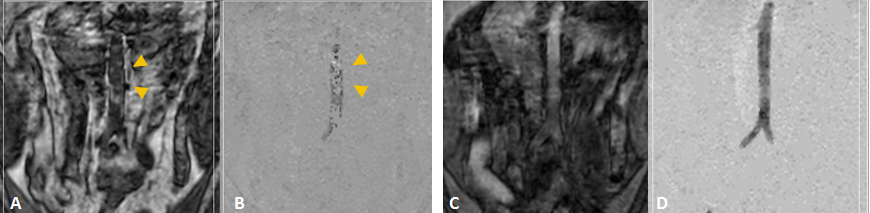
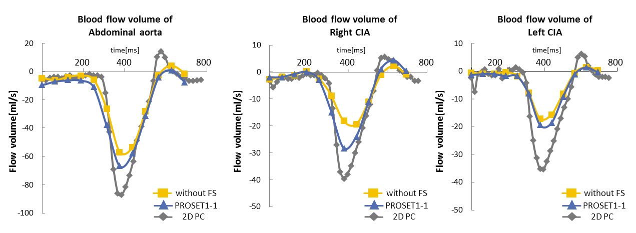
Figure1 shows representative streamline visualizations and representative magnitude and phase images are shown in Fig.2. 4D-PC with ProSet1-1 selective water excitation showed higher SNR compared to without ProSet. The overall blood flow volume with using ProSet1-1 was similar to the 2D reference standard technique (Fig.3).Discussion
Intraperitoneal artifacts and respiration artifacts on the non-suppressed images were observed (Fig.2). Fat suppression technique could reduce these artifacts and improve background suppression. Extending of TR by using ProSet increases SNR. However, excessively extending of TE with ProSet1-2-1 decrease signals. Thus ProSet1-1 is the optimal fat suppression technique to improve vascular visualization.Conclusion
The optimal selective water excitation technique was ProSet1-1 and one could reduce artifacts and provide sensitive hemodynamic sufficiently. Combined use of selective water excitation may be useful for 4D-PC MRI and blood flow visualization of the CIA.Acknowledgements
No acknowledgement found.References
1. Jelena Bock, et al. 4D phase Contrast MRI at 3T:Effect of Standard and Blood-Pool Contrast Agents on SNR, PC-MRA, and Blood Flow Visualization. Magnetic Resonance in Medicine.2010;63:330-338
2. Alex Frydrychowicz, MD, et al. Visualization of Iliac and Proximal Femoral Artery Hemodynamics Using Time-Resolved 3D Phase Contrast MRI at 3T. JOURNAL OF MAGNETIC RESONANCE IMAGING.2007;25:1085-1092
3. Michael Markl, PhD, et al. Time-Resolved 3D MR Velocity Mapping at 3T: Improved Navigator-Gated Assessment of Vascular Anatomy and Blood Flow. JOURNAL OF MAGNETIC RESONANCE IMAGING.2007;25:824-831
Figures