2821
Evaluation of Pediatric Tracheobronchial Anomalies with congenital heart disease using Three-dimensional Turbo Field Echo Magnetic Resonance Imaging Sequence1Radiology, Shanghai Children’s Medical Center affiliated with Shanghai Jiao Tong University school of medicine, Shanghai, People's Republic of China
Synopsis
Tracheobronchial anomalies are common in congenital heart disease (CHD). Cardiovascular anomaly is the principal extrinsic lesion causing tracheobronchial stenosis. MSCT remains an ionizing procedure even though can demonstrate tracheobronchial tree clearly. MRI has the advantage of being non-ionizing and providing excellent soft tissue contrast for the diagnosis of CHD and tracheobronchial anomalies. Spin echo (SE) sequence can demonstrate the tracheobronchial tree but this typically a 2D sequence and therefore difficult to depict the entire tracheobronchial tree optimally. Three-dimensional turbo field echo (3D-TFE) can delineate the entire tracheobronchial tree clearly through post-processing.
Objectives
To evaluate diagnostic accuracy of tracheobronchial anomalies in patients with congenital heart disease using three-dimensional turbo field echo (3D-TFE) magnetic resonance imaging sequence.Materials and Methods
Seventy-five patients with congenital heart disease from December 1, 2013 to December 31, 2015 were retrospectively reviewed. In 75 patients, age ranged from 1.4 months to 134 months; the median age was 10.4 months. Cardiac MR was performed to provide preoperative information about anatomy or/and function. A 3D-TFE and 3D-b-TFE sequences was performed to evaluate tracheobronchial anatomy. All patients also underwent multi-slice computed tomography (MSCT) either before or after MRI. The image quality was evaluated by two reviewers. Inter-modality agreement for tracheobronchial anomaly findings were tested by the kappa coefficient and the sensitivity, specificity of 3D-TFE and 3D-b-TFE for the detection of tracheobronchial anomalies were evaluated.Results
Tracheobronchial anomaly findings on MRI with 3D-TFE in 75 cases were as follow: 31 cases had tracheobronchial stenosis, 9 cases had tracheal bronchus, 3 cases had bridging bronchus, 4 cases had bronchial isomerism and 2 cases had situs inversus bronchus, The predominant causes of tracheobronchial stenosis were double aortic arch, right aortic arch with mirror-image branching, right aortic arch with left aberrant subclavian artery and posterior patent ductus arteriosus (PDA) or ligament, left pulmonary artery sling enlarged left atrium. 3D-TFE provided better image quality as compared to that of 3D-b-TFE There was good inter-modality agreement between 3D-TFE and MSCT for the detection of tracheobronchial anomalies (Kappa0.75). The sensitivity, specificity, accuracy and positive predictive value(PPV) , negative predictive value (NPV), positive likelihood ratio (PLR) and negative liklihood ratio (NLR) of 3D-TFE were 91.3% ,82.8%.88.0%, 89.0%, 85.7%,4.35 and 0.11 respectively.Conclusion
3D-TFE is a useful MRI sequence for demonstrating the tracheobronchial tree and diagnosing tracheobronchial anomalies in congenital heart disease. MRI can supply helpful information for preoperative strategies.Acknowledgements
No acknowledgement found.References
[1] Ming Z, Lin Z Evaluation of tracheal bronchus in Chinese children using multidetector CT. Pediatr Radiol 2007;37:1230-1234. Epub 2007 Oct 2.
[2] Yedururi S, Guillerman RP, Chung T et al. Multimodality imaging of tracheobronchial disorders in children. Radiographics 2008;28:e29. doi: 10.1148/rg.e29.
[3] Lee EY. Advancing CT and MR imaging of the lungs and airways in children: imaging into practice. Pediatr Radiol 2008 Suppl 2:S208-12. doi: 10.1007/s00247-008-0767-3.
[4] Ciet, P., et al., Magnetic resonance imaging in children: common problems and possible solutions for lung and airways imaging. Pediatr Radiol, 2015. 45: 1901-1915. doi: 10.1007/s00247-015-3420-y. Epub 2015 Sep 5.
Figures
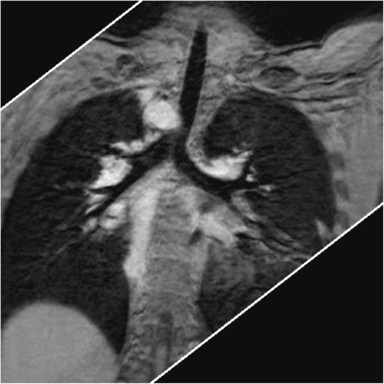
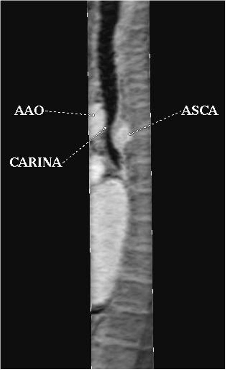
Figure Twelve-year-old girl with corrected transposition of the great artery, Ebstein’s malformation and right aortic arch with aberrant left subclavian artery.
A. Three-dimensional turbo field echo (3D-TFE) coronal minimum intensity projection (MinIP) reconstruction demonstrated prominent stenosis of the lower segment of the trachea.