2819
Estimation of Circulating Blood Volume using Ferumoxytol1National Heart Lung and Blood Institute, National Institutes of Health, Bethesda, MD, United States
Synopsis
Clinical assessment of total blood volume is significant for management of patients with decompensated chronic heart failure. We investigate the feasibility of measuring circulating blood volume as part of an interventional cardiovascular MR exam by measuring T1 changes due to the presence of Ferumoxytol - an intravascular, FDA-approved, iron supplement. Three pigs were scanned prior and twenty minutes post Ferumoxytol administration, from which a mean blood volume of 81.6 ± 1.1 mL/kg was estimated, which approximately overestimates by 15-30% from the literature. This technique has promise as a non-ionizing and non-toxic alternative to measuring patient volume.
Introduction
Cardiovascular MRI can be used to measure parameters such as cardiac function and flow, while minimizing potential exposure to ionizing radiation commonly used for such procedures. However there is not presently a robust method using MRI to measure circulating blood volume, which would inform treatment based on a patient’s volume status. This initial study investigates measuring total blood volume using Ferumoxytol (Feraheme, AMAG, Cambridge, MA, USA), an FDA approved iron-nanoparticle typically used for iron replacement, which has also been investigated as an MR contrast agent due to its long intravascular half-life (~14 hrs) [1-3].Methods
Animal studies were approved by the NHLBI institutional Animal Care and Use Committee. Scanning was performed on a 1.5T Siemens scanner (Siemens Healthcare, Erlangen, Germany), with phased array coils (Body 18) on the anterior and posterior chest. In vitro: Longitudinal relaxavity (r1) of Ferumoxytol was characterized using 7 linearly spaced dilutions between 0.1-1.72 mM in whole blood and T1 measurements were acquired at 37 °C using SASHA [4].
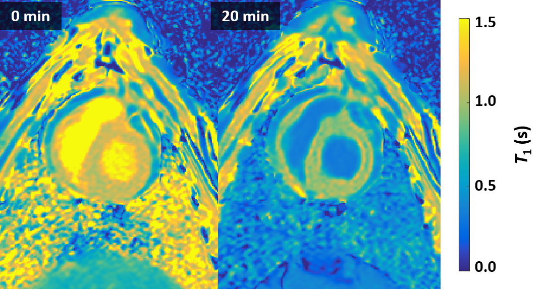
In vivo: SASHA estimations of T1 were acquired breath-held, in a mid-ventricle, short-axis image prior and post administration of 0.6 mg/kg Fe in three pigs (average weight 52 kg). At twenty minutes, a post-administration T1 map was acquired. Total blood volume was estimated using the following relation: TBV = nFe · r1/ ΔR1, where nFe is the molar mass of administered contrast agent, r1 is the relaxivity of Ferumoxytol and ΔR1 is the change (post – pre) in the longitudinal relaxation rate in the blood [5].
Results
SASHA-derived estimation of Ferumoxtyol relaxivity (r1) was 18.0 mM-1·sec-1 ± 0.4 mM-1· sec-1, in good agreement to previous literature [2]. Blood T1 measurements taken within the LV were measured to be 1.46 ± 0.07 s at baseline, which shortened to 0.33 ± 0.01 s at twenty minutes post administration of Ferumoxytol. This yielded an estimated blood volume of 81.6 ± 1.1 mL/kg.Conclusion
Previously reported circulating blood volume for pig species ranges within 56-69 ml/kg [6], which is 69 – 85% of what was measured in vivo using this non-invasive, Ferumoxytol-based technique. These animals were on intravenous fluid, which may contribute to the higher observed values. The technique used here is promising as a MR-derived estimation of circulating blood volume, and can supplement an interventional cardiovascular MR exam. Future experiments will look to validate the blood volume estimated by Ferumoxytol-induced T1 changes.Acknowledgements
This work was supported by the NHLBI DIR (Z01-HL006039, Z01-HL005062).References
[1] S. Reeder, et al. Mathematical Optimization of Contrast Concentration for T1-Weighted Spoiled Gradient Echo Imaging. Magn Reson Med, 2016; 75(4): 1556-1564.[2] C. Corot, et al. Recent advances in iron oxide nanocrystal technology for medical imaging. Advanced Drug Delivery Reviews, 2006; 58: 1471-1504. [3] M. Bashir, et al. Emerging Applications for Ferumoxytol as a Contrast Agent in MRI. JMRI, 2015; 41:884-898. [4] K. Chow, et al. Saturation Recovery Single-Shot Acquisition (SASHA) for Myocardial T1 Mapping. Magn Reson Med, 2014; 71: 2082–2095[5] K. Pannek, et al. Contrast agent derived determination of the total circulating blood volume using magnetic resonance. Magn Reson Mater Phy, 2012; 25: 215-222.[6] https://www.nc3rs.org.uk/blood-sample-volumesFigures