2809
A novel method for Background Suppression in Continuous Arterial Spin Labeling (cASL) AngiographyJianxun Qu1, Bing Wu1, and Zhenyu Zhou1
1MR Research China, GE Healthcare, Shanghai, People's Republic of China
Synopsis
In this work, we implemented background suppression for continuous arterial spin labeling based MR angiography to suppress the noise resulting from the influence of magnetic transfer effect. Pulsed labeling is also incorporated in the background suppression scheme to address the flow void effect in continuous labeling. With the proposed method, the overall vessel clarity was improved without prolonging the scan time.
Purpose
Arterial spin labeling (ASL) angiography with continuous tagging (cASL) features optimal labeling efficiency and being silent [1]. In convention, in the labeling module, the tagging plane is placed underneath the imaging slab so that the cranial arterial flow may be labeled; whereas in the control module, although no tagging plane is needed, a dummy tagging plane is placed above the imaging slab so that the imaging slab may experience same amount of MT effects given the labeling pulse is non-selective. However, in practice only the center of the imaging slab experiences balanced MT effects when the control is subtracted from the tagging, and there tend to be gradual increase of unbalanced MT effects towards the edge of the imaging slab. The so formed background noise might deteriorate the angiogram, especially for distal arteries near the tagging plane. In this work, we propose novel background suppression method to reduce this effect.Methods
The pulse sequences of the proposed method as well as the current approach are shown in Fig.1. The background suppression was achieved via two hyper-secant adiabatic pulses, with the first one inserted in the middle of continuous tagging and the second one placed right after the continuous tagging. After the first inversion pulse is played out, the continuous tagging resumes immediately after. Thus the magnetic transfer effect was exerted with the same magnitude and opposite frequency for the same time duration. A short delay time was waited before the following zero TE acquisition. All the adiabatic inversion slab was placed concentrically to the imaging slab. The thickness of the first inversion slab covers the region confined with the paired continuous tagging planes. The second inversion pulse has the inversion thickness used in flow sensitive alternating inversion recovery (FAIR) to invert blood about to enter the imaging slab during readout segment. Quadrature-phase saturation pulse with phase cycling was played twice prior to continuous tagging to eliminate any residue signal. The proposed method was implemented on a 3.0T whole body system (GE, Discovery 750W, Milwaukee, Wisconsin) equipped with a 24 channel head coil. For comparison, the background suppressed sequence with the thickness of second inversion pulse set to the same value for labeling and control was also implemented to exclude the influence of FAIR tagging. A healthy volunteer was recruited with consent form acquired, and cASL angiography scans with and without background suppression were performed. The total tagging preparation duration was 1650ms for both ASL-MRA with and without BGS modulation. In BGS sequence, the continuous tagging was 1600 ms and the waiting time was 50 ms. The zTE imaging parameters are listed as follows: FOV 180 mm, isotropic resolution 1.2 mm, spokes per segment 512, and flip angle 4 degree. The continuous tagging plane was 20 mm away from imaging volume. The thickness of the first inversion slab was 220 mm. The thickness for the second inversion slab was 220 mm and 320 mm for control and labeling group respectively. Background noise and artery intensity level were quantitatively compared.Results
Fig.2 shows the maximum intensity projection (MIP) angiogram from ASL-MRA without BGS (Fig.2a), with BGS and the same thickness for second inversion pulse (Fig.2b) and with BGS and FAIR tagging scheme for the second inversion pulse (Fig.2c). Heavy MT effect induced background noises could be observed in purely continuous tagging MRA (Red Round Box, Fig.2a). Arteries indicated by white arrow have conspicuous better visualization in both BGS MRA. Part of anterior cerebral artery (ACA) could only be seen in BGS modulated MRA but not in conventional continuous tagging. The background noise level at upper and middle slices (Blue dot), MIP signal at left middle cerebral artery (LMCA) and left internal cerebral artery (LICA) are compared in Fig.3. It can be seen that using modulated BGS led to higher level of vascular signal as well as more homogenous background noise level.Discussion and Conclusion
In this work, a novel method of background suppression with cASL tagging method was proposed. It effectively eliminated the non-uniform noise distribution due to MT effects in cASL. Not only it improves the overall clarity of the resulting angiogram, it helps to visualize the distal vessels often obscured by high noise level. This method does not prolong the scan time and can be readily implemented into current cASL MRA acquisition. An area of work needs to be addressed in the future is the optimization of the inversion time and post labeling time.Acknowledgements
No acknowledgement found.References
[1] Y.Fujiwara, Improving in visualization of carotid artery uniformity using silent MRA, Radio Phys Technol, 2016.Figures
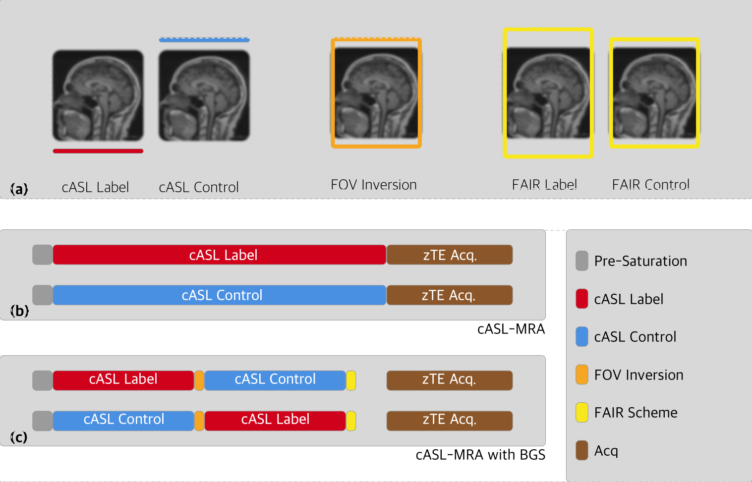
Fig.1 Pulse sequence of continuous without background suppression (BGS) (b) and with BGS (c). The continuous ASL pulse and inversion pulse position are presented in (a).
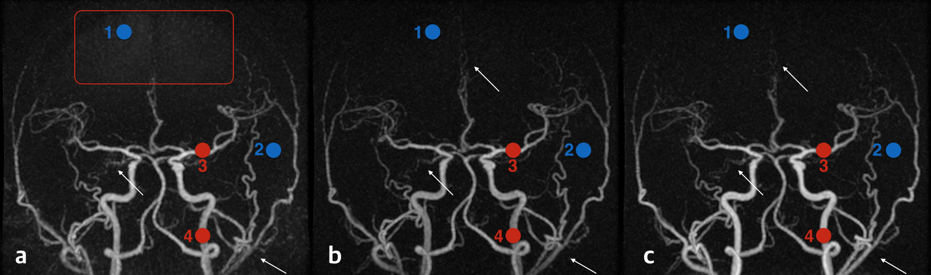
Fig.2 MRA results: cASL labeling without BGS (a), ASL with BGS where the second inversion pulse used the same thickness for labeling and control (b), and ASL with BGS where the second inversion took FAIR scheme (c).
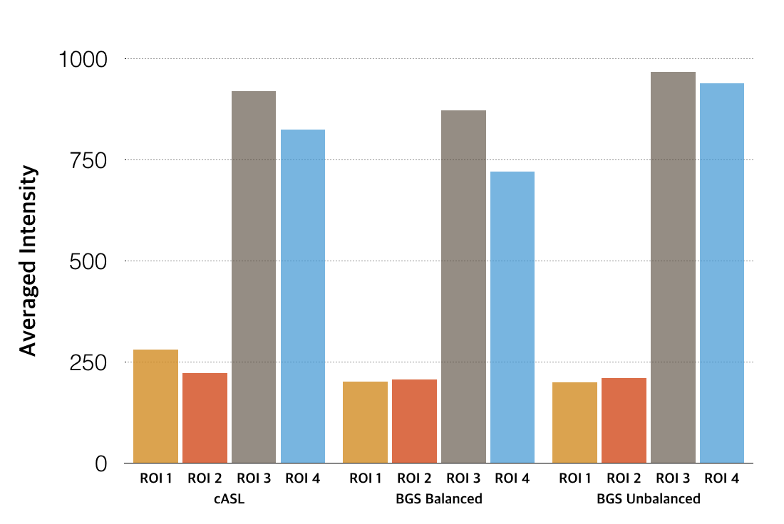
Fig.3 Signal intensity of left ICA, left MCA, and background noise.