2802
Novel ASL-based flow imaging for cardiovascular hemodynamics and valvular function visualization: comparison with a feasibility study1Department of Radiological services, Tokyo Women’s Medical University Hospital, Tokyo, Japan, 2Department of Diagnostic Imaging & Nuclear Medicine, Tokyo Women’s Medical University Hospital, Tokyo, Japan, 3Philips Electronics Japan, Tokyo, Japan
Synopsis
We developed a novel ASL-based flow imaging (ASL-Flow) with modified flow-sensitive alternating inversion recovery technique and Look-Locker sequence to evaluate cardiovascular hemodynamics and valvular dysfunction. We validate that time-intensity curves of pulmonary artery form ASL-Flow matches for time-velocity curves from 2D phase-contrast MRI. ASL-Flow can visualize more clearly regurgitateion and stenotic jet in valvular diseases than standard cine imaging. The scan time for ASL-Flow is short (15 seconds/slice), and the image reconstruction needs no special software; therefore, this is easy to clinically use. We promise that ASL-Flow is a feasible non-contrast imaging to detect hemodynamics abnormality in structural heart disease.
Purpose
In structural heart disease (SHD), the assessments of cardiovascular hemodynamics and valvular function are clinically important for the determination of therapeutic option. Commonly, such information is obtained by phase-contrast (PC) imaging and 2D cine MRI technique. PC sequence is established as the assessment of cardiovascular hemodynamics (1, 2); however, the accurate assessment using PC MRI potentially depends on radiologist’s skill. Four-dimensional (4D) flow MRI has recently been reported as a unique non-invasive flow imaging (3), whereas a special sequence and dedicated software are required. Moreover, data acquisition and post-processing analysis of 4D flow MRI are time-consuming. Therefore, a clinical usage is still limited. It is well known that conventional 2D cine MRI with steady state free precession is useful for the assessment of cardiovascular hemodynamic (4.5), and provides insight to valvular dysfunction (6). Recently, non-contrast flow imaging using 3-Tesla MRI and arterial spin labeling (ASL) have an important role for the analysis of cerebral blood flow. However, the ASL flow imaging is not applied for cardiac MRI due to heart beat and the complex flow in the four chamber of the heart. The present study is to propose a novel ASL-based flow imaging (ASL-Flow) with modified flow-sensitive alternating inversion recovery (FAIR) technique and Look-Locker sequence to evaluate cardiovascular hemodynamics and valvular dysfunction, and to investigate the optimal turbo field echo (TFE) pre-pulse slice thickness in ASL.Methods
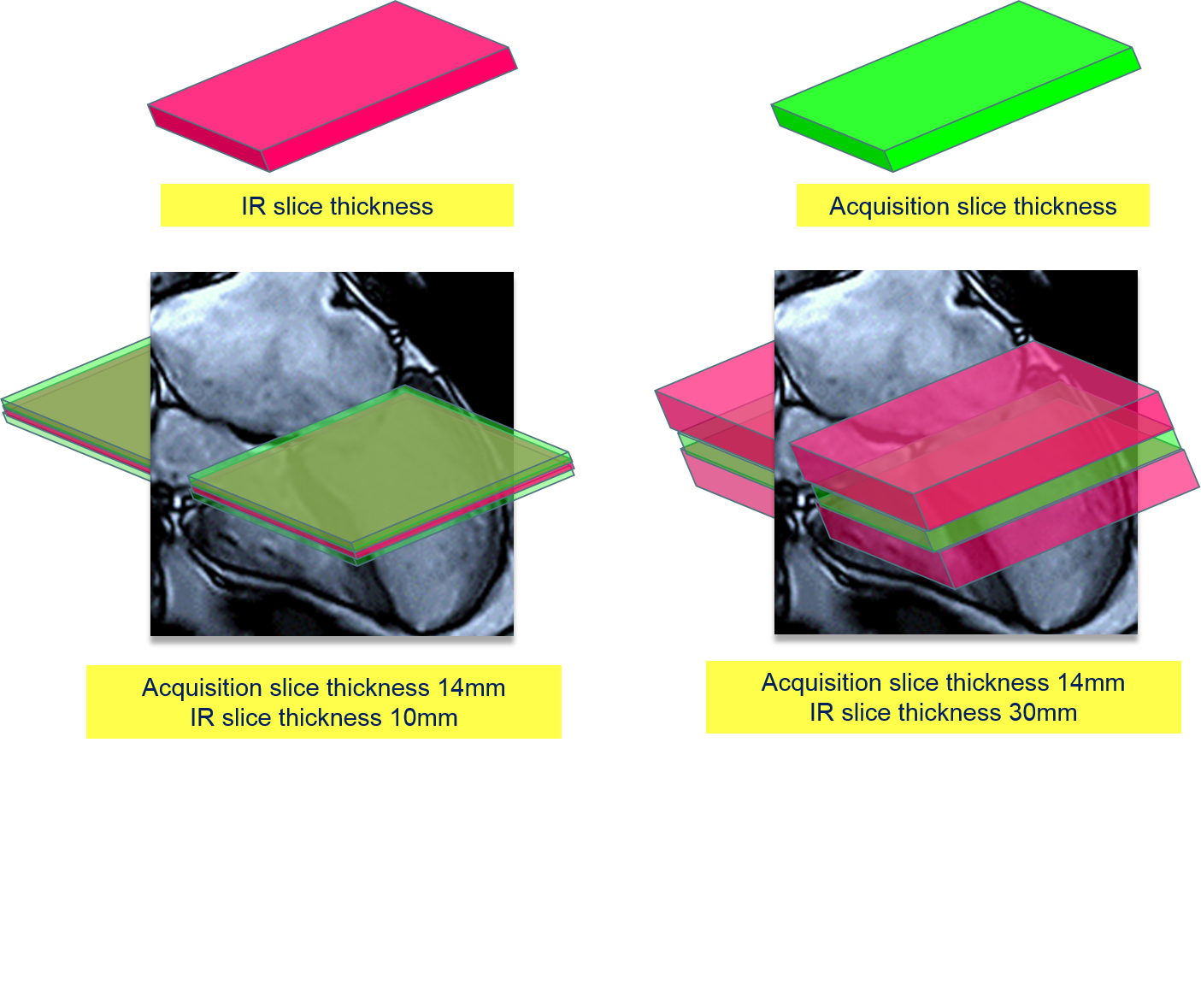
[Subjects] For six male healthy volunteers (age range: 28-41 years), ASL-Flow (Fig. 1) and PC MRI with 3.0T MR unit (Ingenia, Philips healthcare) were performed. We hypothesize that the inflow effect depends on the thickness of slice-selective inversion-recovery (SSIR) labeling pulse in the ASL-flow sequence. First, the optimal thickness of SSIR labeling pulse to maximum the inflow was examined. Second, time-intensity curves from ASL-Flow for aorta, pulmonary artery, and inferior vena cava were compared with time-velocity curves from standard PC MRI. Finally, we investigate the diagnostic ability of valvular function in patients SHD, compared with standard 2D cine MRI. [ASL-Flow] Image parameters: slice thickness=14mm, FOV 180mm, NSA=1, pixel size=2.81X2.37mm², TR=20msec, acquisition time=15sec, number of heart phase=44.Results&Discussion
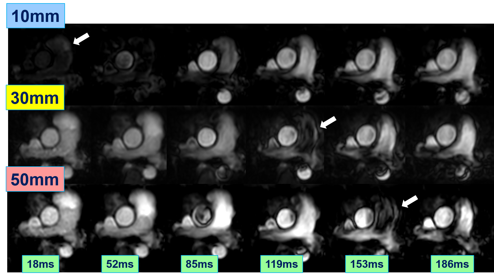
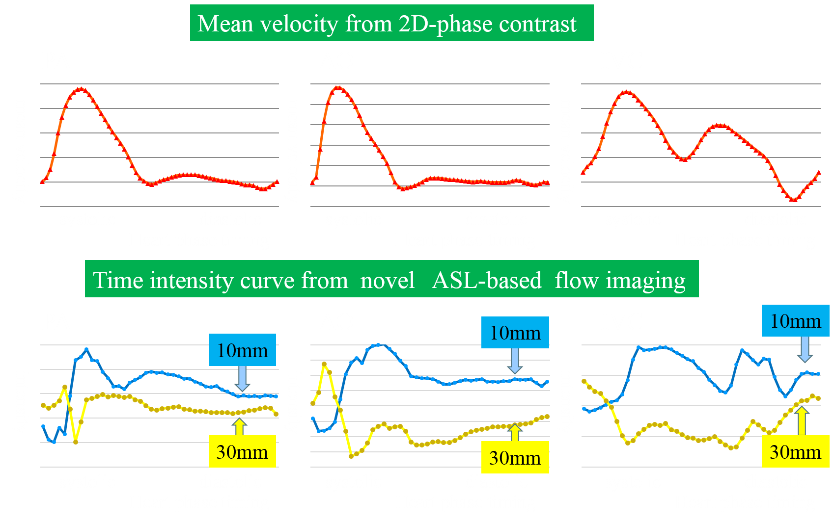
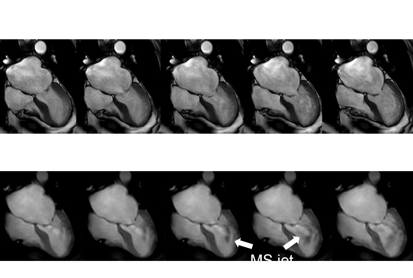
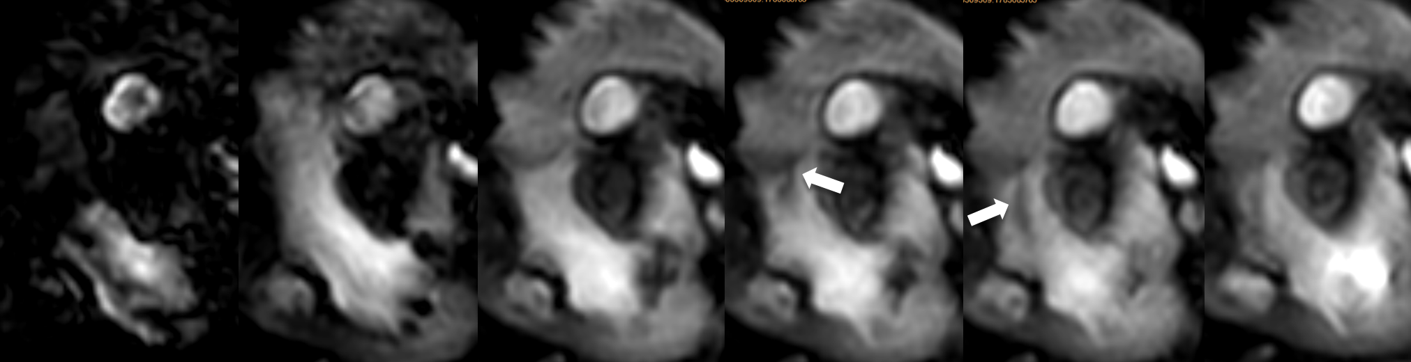
Flow imaging with thickness of SSIR labeling pulse of 10mm shows black tags in pulmonary arteries at initial images of early systole. The following images throughout a cardiac cycle can visualize continuous flow. Flow imaging with thickness of 30 and 50mm appears transient black tags at mid-systole; therefore, pulmonary arterial flow was interrupted (Fig. 2). Time-intensity curves from 10mm thickness of ASL-Flow broadly matched for time-velocity curves from PC MRI (Fig. 3). On the other hand, time-intensity curves from 30mm thickness showed a declining at the peak time of time-velocity curves from PC MRI. In a case of mitral stenosis who cannot be detected by standard cine imaging, ASL-Flow can visualize a clear jet flow due to stenotic valve at diastole (Fig. 4). In a case of repaired complete transition of great arteries, aortic regurgitation could be diagnosed using ASL-Flow (Fig. 5). Standard cine imaging could assess due to artifact of post-operative material. We focused on developing a simple, easy and user-friendly non-contrast flow technique, which is expected for clinical contributions.Conclusion
ASL-Flow using a thickness of SSIR labeling pulse of 10mm was well correlated with the flow data from PC MRI. This sequence is a feasible non-contrast flow imaging to detect hemodynamics abnormality in SHD and easy to clinical use.Acknowledgements
No acknowledgement found.References
(1): Powell AJ, Geva T. Blood flow measurement by magnetic resonance imaging in congenital heart disease. Pediatr Cardiol. 2000 Jan-Feb;21(1):47-58.
(2): Powell AJ, Maier SE, Chung T, et al. Phase-velocity cine magnetic resonance imaging measurement of pulsatile blood flow in children and young adults: in vitro and in vivo validation. Pediatr Cardiol. 2000 Mar-Apr;21(2):104-10.
(3): Rose M, Rahman O, Schnell S, et al. 4D flow MRI demonstrates changes in cardiovascular haemodynamics in complex congenital heart disease. Eur Heart J Cardiovasc Imaging. 2016 Sep 27. pii: jew204. [Epub ahead of print]
(4): Maceira AM, Prasad SK, Khan M, et al. Normalized left ventricular systolic and diastolic function by steady state free precession cardiovascular magnetic resonance. J Cardiovasc Magn Reson. 2006;8(3):417-26.
(5): Sakamoto I, Takemoto M, Nagao M, et al. Giant pulsatile coronary aneurysms associated with pulmonary atresia with an intact ventricular septum after a Fontan procedure. Circulation. 2014 Sep 30;130(14):e121-3.
(6): Krombach GA, Kühl H, Bücker A, et al. Cine MR imaging of heart valve dysfunction with segmented true fast imaging with steady state free precession. J Magn Reson Imaging. 2004 Jan;19(1):59-67.
Figures