2798
Diagnostic Accuracy Of 3D Head And Neck Joint Black-Blood Vessel Wall Imaging In Patients With Carotid Artery StenosisComparison with DSAZhenjia Wang1, liu wen2, and yu wei3
1Department of Radiology, Department of Radiology, Anzhen Hospital, CapitalMedical University, Beijing, China, beijing, People's Republic of China, 2Department of Radiology, Anzhen Hospital, CapitalMedical University, Beijing, China, 3Department of Radiology, Anzhen Hospital, CapitalMedical University, Beijing, China, beijing, People's Republic of China
Synopsis
Joint head and neck vessel wall imaging technology with variable flip angle turbo spin echo (SPACE)1 has recently been introduced as a promising MRI method for simultaneous evaluation of extra-cranial and intra-cranial vessel wall. However, this technique is yet to be validated with established imaging techniques. We aim to evaluate the accuracy of this technique using DSA as reference in assessing carotid artery stenosis and plaque morphology.
Purpose
Joint head and neck vessel wall imaging technology with variable flip angle turbo spin echo (SPACE)1 has recently been introduced as a promising MRI method for simultaneous evaluation of extra-cranial and intra-cranial vessel wall. However, this technique is yet to be validated with established imaging techniques. We aim to evaluate the accuracy of this technique using DSA as reference in assessing carotid artery stenosis and plaque morphologyMethods
Eleven patients with carotid artery stenosis who were scheduled to undergo carotid artery stent were studied with MR imaging within 1 week of the procedure. Patients were imaged with a phased-array carotid surface coil and head coil on a 3.0-T Scanner (Siemens Verio). 3D head and neck joint black-blood vessel wall Imaging was performed with 0.625mm isotopic resolution. Internal carotid artery from extra-cranial to the cranial was divided to 7 segments (C1-C7) by Bouthillier classification. The diameter of each ICA segment, proximal and distal lumen of stenosis, minimal luminal diameter were measured. The degree of the stenosis was calculated according to the North American Symptomatic Carotid Endarterectomy(NASCET) criteria. To check the accuracy of our data and eliminate mismatch between different techniques, we compared the MR images with DSA scans of the same patients from the same orientation. The paired samples T-test and Pearson correlation was used to evaluate the agreement between MR and DSA measurements.Result
A total of 11 patients, 22 arteries, 12 plaques, 190 measurements were included in this study. Among 12 plaques, 7 plaques were located in carotid bifurcation, 2 were located between C1-C2 , 3 plaques were located in the C3-C5. There are 4 plaques shown as hyper-intensity, 8 shown iso- to hypo-intensity. MR imaging showed excellent agreement (intraclass correlation coefficient [Is intraclass correlation or pearson correlation which ismentioned in methods.]0.937) with DSA on luminal diameter of each segment and stenosis. There are no significant difference in the measurements from C1-C3 segments between MRI and DSA. Carotid artery stenosis was accurately diagnosed fromsegment 1 to segment 3 of ICA in MRI compared with DSA.Discussions
There were significant differences in the measurements from segment 3 to segment 7 of ICA. The main reason is the cervical part ascends almost vertically to the base of the skull where it turns into the carotid canal in the petrous part of the temporal bone and the intracranial internal carotid artery vessel usually growth shape distortion, so that the data of segment 3 to segment 7 of ICA in MRI could not match well with DSA in same patient.Conclusion
Head and neck joint black-blood vessel wall Imaging can accurately and quantitatively assess arterial morphology to evaluate lumen stenosis, especially in the segment 1 to segment 2 of ICA.Acknowledgements
No acknowledgement found.References
1 Xie, Y. et al. Improved black-blood imaging using DANTE-SPACE for simultaneous carotid and intracranial vessel wall evaluation. Magnetic resonance in medicine : official journal of the Society of Magnetic Resonance in Medicine / Society of Magnetic Resonance in Medicine75, 2286-2294, doi:10.1002/mrm.25785 (2016).Figures
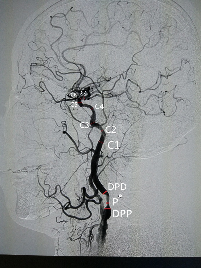
Mearsuments on DSA and MRI
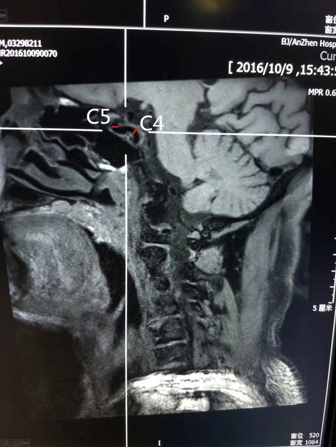
Mearsuments on DSA and MRI
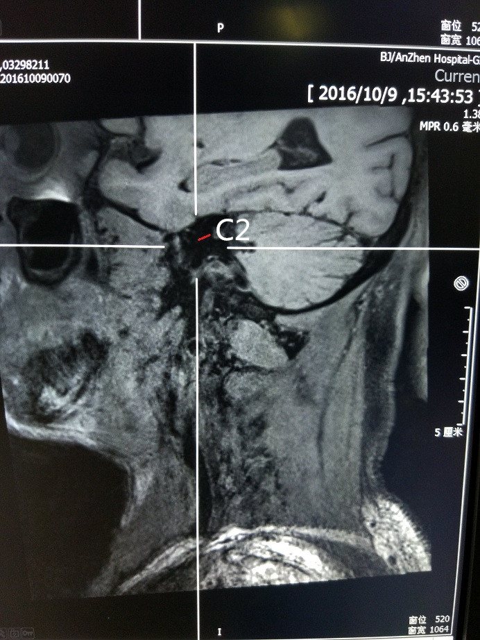
Mearsuments on DSA and MRI
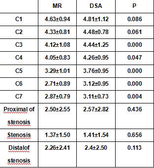
Comparison between DSA and MR measurements of luminal diameters (Mean±SD, mm)