2787
Optimization of 3D black-blood multi-echo T2* weighted sequence in carotid artery1Department of Radiology, University of Cambridge, Cambridge, United Kingdom, 2Department of Radiology, Cambridge University Hospitals NHS Foundation Trust, Cambridge, United Kingdom
Synopsis
Carotid T2* weighted images have multiple applications, including differentiating plaque components and detecting ultrasmall superparamagnetic iron oxide (USPIO) contrast agents. The current work develops and
Purpose
The purpose of this study is to develop and optimize a 3D black-blood multi-echo T2* weighted sequence for carotid imaging, through optimal blood suppression and dedicated k-space ordering.Methods
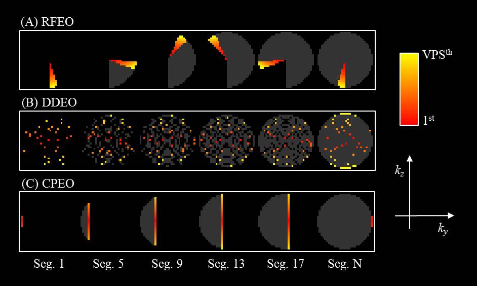
Sequence: Two blood suppression techniques, iMSDE1 and DANTE2, were implemented into a 3D multi-echo fast spoiled gradient echo sequence (FSPGR). A segmented multi-shot acquisition was used to acquire six echoes during the readout. The parameters for the readout were: views-per-segment (VPS)=40; delay time after each segment (TD)=600ms; TE/TR=4.9-31.8/36.8ms with echo spacing=5.4ms; flip angle=15°; receiver bandwidth=±31.25kHz; Acquisition matrix, resolution and orientation: 224×224×40, 0.6×0.6×1.4mm3, axial plane. In the readout, three different k-space orderings were implemented and compared. The first method was a Radial Fan-beam Encoding Ordering (RFEO3, Figure 1A). In this method, the k-space points in the ky-kz plane were first sorted into different segments by the polar angles, and then by their distance from the k-space centre. Blood suppression was applied at the beginning of each segment. The second method was the Distance-Determined Encoding Ordering (DDEO, Figure 1B). In this method, all the k-space points in the ky-kz plane were sorted by their distance from the k-space centre, and then the ith×N points were allocated to the ith place in each of the segment (i from 1 to VPS, N is the number of segments). Blood suppression was again applied at the beginning of each segment. The third method was the Centric Phase Encoding Order (CPEO, Figure 1C), where each of the segments acquires only a single line along kz in the k-space. Blood suppression was applied at the beginning of each segment.
Bloch simulations: Bloch equation simulations were performed to study the signal evolution of the vessel wall with the different blood suppression methods and k-space orderings. The relaxation times used in the simulation were: T1_blood=1500ms, T2_blood=128ms, T1_wall=1000ms and T2_wall=50ms. Other parameters were the same as the volunteer protocol.
Volunteer scans: Five volunteers were recruited for this study. The volunteer experiments were conducted under a research ethics agreement and all volunteers gave informed written consent. All the scans were performed using a 3T system (MR750, GE Healthcare, Waukesha, WI), using a 4 channel phased-array neck coil (PACC, MachNet, Roden, The Netherlands). The scanning time for the RFEO and DDEO acquisitions was 6min15s, and 7minutes for CPEO. Lumen and wall contours of three contiguous slices 5mm below the bifurcation were manually drawn by an experienced reviewer in OsiriX (OsiriX 5.5.2, Pixmeo, Geneva, Switzerland). Vessel wall signal-noise-ratio (SNR) and wall-lumen contrast-noise-ratio (CNR) were calculated. The data was presented as mean±sd (standard deviation). A paired student’s t-test was used to compare the difference. The statistical analysis was performed using R (version 3.2.2).
Results
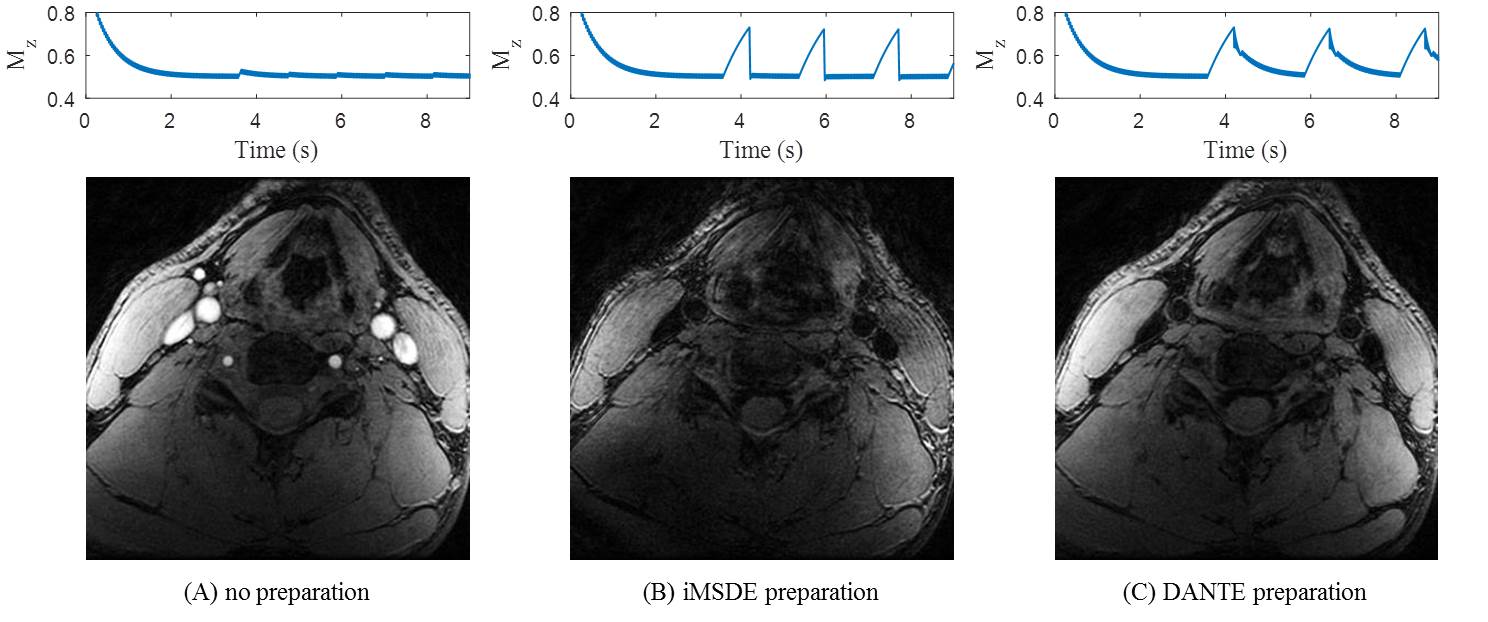
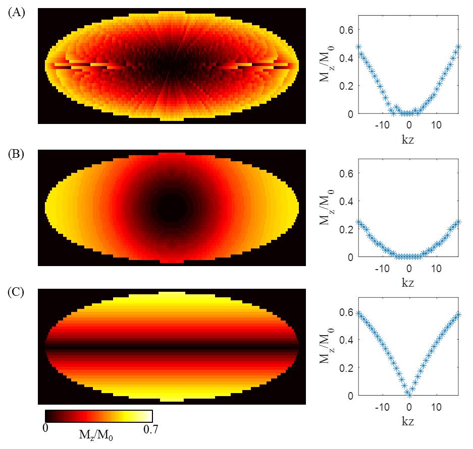
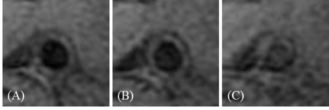
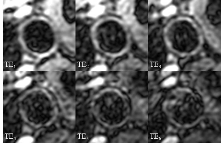
To achieve adequate blood suppression, the first moment for iMSDE preparation was 6666.7 mTms2/m, and the optimal DANTE parameters were: number of pulses 150, flip angle 13˚, repetition time ~1ms. Figure 2 shows the Bloch simulation of the change of vessel wall longitudinal magnetization over time and the corresponding volunteer images using the different blood suppression methods. The readout was using RFEO method. The DANTE preparation has a more stable signal across the image, and has higher wall SNR compared to the iMSDE preparation (18.1±6.6 vs. 15.3±7.4, p=0.02). Figure 3 shows the Bloch simulation of the longitudinal magnetization of blood using the different k-space orderings. Since the signals in the k-space centre determine the contrast in the image domain, and the image signal is proportional to the longitudinal magnetization, a method that could allocate the low Mz points in the k-space centre region achieves better blood suppression. Under this assumption, the DDEO (B) method should achieve the best blood suppression. Figure 4 shows an example of a DANTE prepared sequences with the three different k-space orderings. RFEO and DDEO achieved better blood suppression than CPEO method. The results show that volunteer wall-lumen CNR of RFEO (12.2±4.7) and DDEO (10.6±6.0) are comparable, and they are both higher than the CPEO method (8.6±6.1). However, none of the differences was significant (p>0.05). Figure 5 shows an example of DANTE prepared multi-echo T2* weighted images using RFEO method as readout.Discussion and conclusions
This study developed and optimized a 3D black-blood multi-echo T2* weighted sequence through Bloch simulation and volunteer scans. The results show that the DANTE preparation is better than iMSDE in terms of image SNR. In addition, the RFEO and DDEO k-space ordering methods have similar blood suppression efficiency, and are both better than the CPEO method.Acknowledgements
We acknowledge funding from NIHR BRC and Addenbrooke's Charitable Trust.References
1. Wang J, Yarnykh VL, Yuan C. Enhanced image quality in black-blood MRI using the improved motion-sensitized driven-equilibrium (iMSDE) sequence. Journal of Magnetic Resonance Imaging. 2010 May 1;31(5):1256-63.
2. Li L, Miller KL, Jezzard P. DANTE: prepared pulse trains: A novel approach to motion-sensitized and motion-suppressed quantitative magnetic resonance imaging. Magnetic resonance in medicine. 2012 Nov 1;68(5):1423-38.
3. Vu AT, inventor; General Electric Company, assignee. Method and apparatus for acquiring MR data with a segmented multi-shot radial fan beam encoding order. United States patent US 7,265,547. 2007 Sep 4.
Figures