2783
Improvement of fat separation for simultaneous carotid and intracranial vessel wall imaging by dual-echo fat-water imaging1Shenzhen Institutes of Advanced Technology, Chinese Academy of Sciences, Shenzhen, People's Republic of China
Synopsis
In this study, we used dual-echo fat-water imaging technique in variable flip angle turbo spin echo sequence for simultaneous carotid and intracranial artery imaging to obtain fat-free vessel wall images. Results show that the technique was promising for simultaneous carotid and intracranial vessel wall imaging to improve the SNR and better delineation of carotid vessel wall.
Introduction
Magnetic resonance vessel wall imaging (VMI) has emerged as a promising noninvasive modality for characterizing atherosclerotic plaque in carotid and intracranial arteries [1]. Recently, high resolution 3D isotropic black-blood sequence using variable flip angle turbo spin echo (SPACE) at 3T was developed to simultaneously evaluate the carotid and intracranial arterial vessel walls [2-3]. Vessel wall imaging would be benefited from good fat suppression as the bright fat signal would obscure the observation the outer boundary of vessel wall. Traditionally, fat saturation was used for fat suppression. However, for simultaneous carotid and intracranial VWI, the spectrally-selective technique fails to saturate fat completely due to the field inhomogeneity in large imaging volume, and even results in the decreased SNR in cervical region. In this study, we used dual-echo fat-water imaging technique in carotid and intracranial VWI to obtain fat-free images.Material and methods
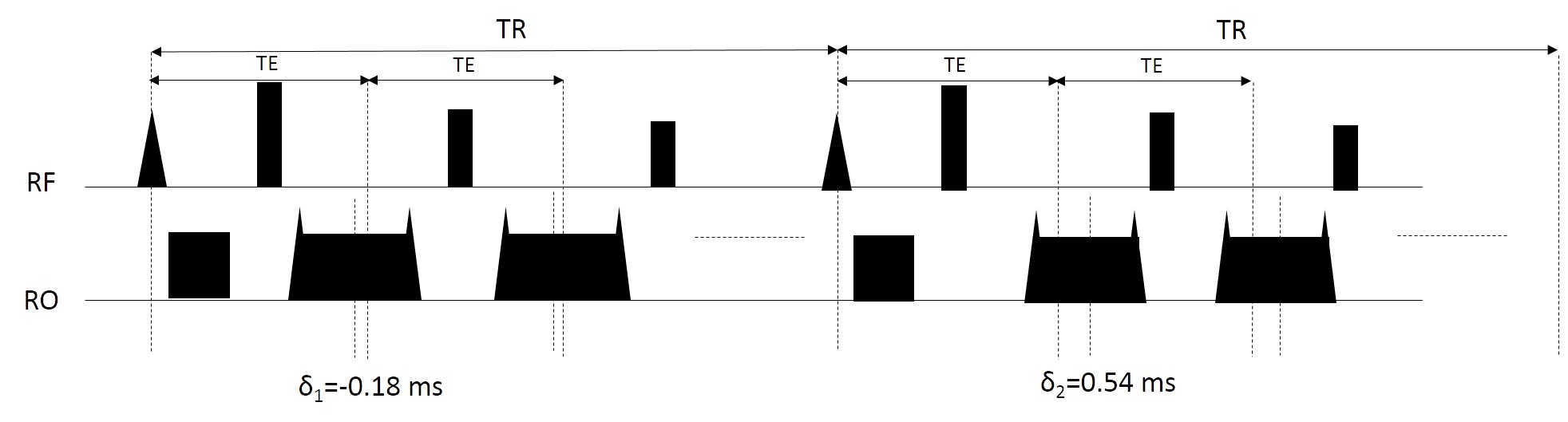
All experiments were conducted in a Siemens 3T system (TIM TRIO) with an elaborated 32-channel head/neck coil [4]. The original SPACE sequence was slightly adapted to fat-water imaging (fw-SPACE). The center of echoes were shifted -0.18 ms and 0.54 ms away from the center of refocusing pulses in two successive acquisitions (Figure 1), which corresponded to -π/6 and π/2 phase differences between water and fat at 3T. The shifted echo time increases the echo spacing by 1.08 ms. The imaging slab was set in sagittal view covering whole brain and neck. Other imaging sequence parameters were: FOV = 188 mm, imaging matrix = 256 * 256 * 192, resolution = 0.73 mm isotropic, TR/TE = 700/11 ms, echo train length = 47, bandwidth = 781 Hz/pixel, echo spacing = 4.68 ms, GRAPPA acceleration factor = 2 in phase encoding direction. The total acquisition time was 13 min 26 sec for fw-SPACE.
The volunteer study was then performed using both fw-SPACE described above and original SPACE with fat saturation (fs-SPACE) for comparison. The volunteer study was IRB approved. Four healthy young volunteers were recruited with all informed consents. The parameters for fs-SPACE were the same as above except that two averages were used to keep the same scan time as the fw-SPACE.
Fat water separation with projected power method [5] was applied to the dual echo data to obtain the water-only and fat-only images. SNRs in the intracranial and extracranial regions were then compared in the water-only images and fat-saturated images.
Results
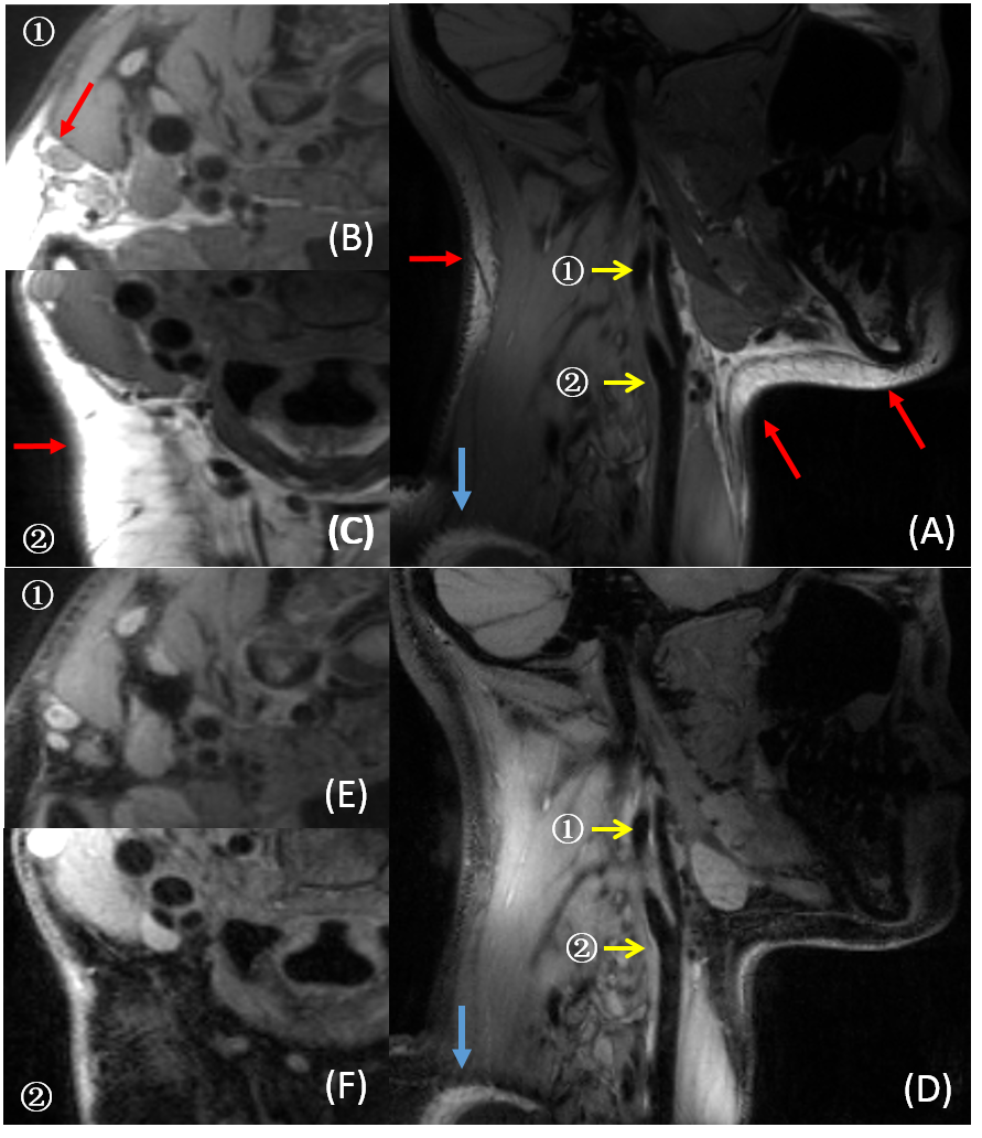
Figure 2 shows the images from fat-saturated images (A-C) and water-only images (D-F). All images are shown in the same window levels. Incomplete fat saturation were observed in all volunteers, especially in the cervical regions (read arrows). In water-only images, the fat signal was uniformly suppressed in the region of interest. Additionally, the water-only images show better SNR in the neck area than the fat-saturated images, as the water signals are partially saturated due to the large field inhomogeneity in the neck area. Most interestingly, the contrast of vessel wall to background is improved due to the suppression of intramuscular fat in water-only images (see the inserts).
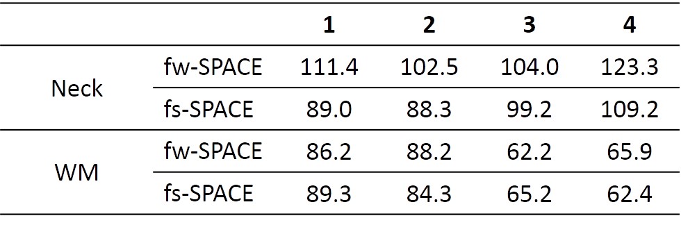
Table 1 compares the SNR between fw-SPACE and fs-SPACE in muscle around neck and white matter. The SNRs are obviously improved in the neck area (from 5%~30%), while the SNRs in white matter are almost identical.
Discussion and conclusions
In our study, the dual-echo was chosen as [-π/6, π/3], which is not optimal for fat water separation. However, the optimal scheme [0, π] would largely increase the echo spacing by 2.16 ms at 3T, which would compromise the SNR and prolong the acquisition.
The SNR improvement in neck area varies in different volunteers, which could be attributed to different achievable shimming performances in different volunteers. However, in white matter, the SNRs are almost the comparable in the two sequences. This could be expected as the fat saturation has little effect in brain where field shift is small. Meanwhile, the effective number of signal averages of the fat-water imaging approximates to the number of averages of fs-SPACE used [6].
The limitation of the technique is the doubled scan time, which may be further improved by acquiring dual-echo signal with reverted readout gradient in one echo spacing. Partial echo acquisition can be incorporated to reduce the echo spacing.
In conclusion, SPACE with fat-water imaging was promising for simultaneous carotid and intracranial vessel wall imaging to improve the SNR and better delineation of carotid vessel wall.
Acknowledgements
This research was supported by the National Natural Science Foundation of China No. 81327801, 61302040, 11504401, 81527901, 81301242, U1301258, National Key R&D Program (Grant No. 2016YFC0100100), Shenzhen Science and Technology Research Program No. JCYJ20150630114942317 and JCYJ20150521094519487References
[1] Yuan C, Oikawa M, Miller Z, Hatsukami T. MRI of carotid atherosclerosis. J Nucl Cardiol 2008;15:266–275.
[2] Zhang L, Tao YJ, Hu XQ, Wu J, Liu X, and Chung Yiu-Cho. High resolution three dimensional imaging of extracranial and intracranial arteries. In Proc. Intl. Soc. Mag. Reson. Med. 23, 2015, p.0552
[3] Xie Y, Yang Q, Xie G, Pang J, Fan Z, Li D. Improved Black-blood Imaging Using DANTE-SPACE for Simultaneous Carotid and Intracranial Vessel Wall Evaluation, Magnetic Resonance in Medicine, 2015; doi: 10.1002/mrm.25785
[4]Hu X, Li Y, Zhang L, Zhang XL, Liu X, Chung Yiu-Cho. A 32-channel coil system for MR vessel wall imaging of intracranial and extracranial arteries at 3T, Magnetic Resonance Imaging 2016; doi: 10.1016/j.mri.2016.10.018
[5] Zhang T, Chen Y, Bao S, Alley MT, Pauly JM, Hargreaves BA and Vasanawala SS. Resolving phase ambiguity in dual-echo dixon imaging using a projected power method, 2016, Magn. Reson. Med.. doi:10.1002/mrm.26287
[6] Xiang QS, Two-point water-fat imaging with partially-opposed-phase (POP) acquisition: an asymmetric dixon method. Magnetic Resonance in Medicine 2006, 56:572-584
Figures