2778
3D visualization of Vascular Cell Adhesion Molecule-1 (VCAM-1) specific Ultrasmall Superparamagnetic Iron Oxide (USPIO) nanoparticles in the atherosclerotic mouse with accelerated self-navigated radial 4D-MRI.1Medizinische Klinik und Poliklinik I, Universitätsklinikum Würzburg, Würzburg, Germany, 2Experimentelle Physik V, Universität Würzburg, Würzburg, Germany, 3Institut für Diagnostische und Interventionelle Neuroradiologie, Universitätsklinikum Würzburg, Würzburg, Germany, 4Fraunhofer IIS, Fraunhofer EZRT, Magnetresonanz- und Röntgenbildgebung (MRB), Würzburg, Germany
Synopsis
USPIO-based, functionalized contrast agents targeting VCAM-1 enable the visualization of inflamed areas in the vessel wall during early atherogenesis. We present a novel 3D technique for coverage of the whole aortic arch at high spatial resolution and a time-resolved detection of nanoparticles with an ECG-free flow-compensated radial 3D-Cine acquisition. T2* weighted 3D-Cines were acquired using two different gradient echoes to provide phase difference maps. The results indicate a reliable detection of the nanoparticles with the new method due to a true 3D coverage of the aorta and the additional phase information.
Purpose
VCAM-1 is known to play a critical role in early atherogenesis. It is expressed highly on activated endothelial cells and mediates the recruitment and adhesion of immune cells to vascular inflammation sites. The use of functionalized contrast agents targeting VCAM-1 enables the visualization of these inflammation areas during the development of atherosclerosis, since the USPIOs accumulated in the inflamed vessel wall lead to a signal loss in T2* weighted images1. Conventional methods to visualize these signal cancelations are based on triggered FLASH measurements and are usually limited to 2D-measurements at few previously selected locations along the aorta, making a whole coverage of the vessel unfeasible. A further problem of 2D measurements is the usually large slice thickness, since objects with large susceptibility differences outside the image slice (e.g. lung tissue) can lead to misleading signal voids in the 2D image. In this abstract we present a new method for detection of USPIOs, which is based on a ECG-free radial multi-echo 3D-Cine measurement. This new approach provides an accelerated coverage of the complete aortic arch and supplemental information from the acquired phase difference maps.Materials and Methods
Animal Handling
ApoE-/- mice (age: 12 weeks) were anesthetized with isoflurane (1.5-2% in oxygen) and kept on a constant body temperature of 37°C. MR measurements were performed at the aortic arch on two subsequent days. After the baseline measurement on day 1, the contrast agent Polyethylene glycol (PEG)-USPIO-VCAM-1 peptide (P03011, Guerbet Research, France) was applied (concentration: 600 µmol/kg) and the measurements were repeated 24 hours after injection of the contrast agent.
MR measurements
MR imaging was performed on a 17.6T small animal MRI scanner with a 24 mm birdcage coil and a 1 T/m gradient system. T2* weighted 3D-Cines were acquired with a flow compensated radial gradient echo sequence. Self-navigation signals were extracted from the radial DC signal and used for a retrospective Cine reconstruction, as described previously2,3.The scan parameters were: TR=4.0 ms, FOV: 25x25x5 mm3, spatial resolution: 98x98x98 µm3, 20 Cine frames. Data was acquired at two different echo times, TE1=1.5 ms, TE2=2.3 ms. The total scan time for both data sets was 25 minutes. All reconstructions were performed with MATLAB (The Mathworks, Inc, USA). 2D slices were extracted from the reconstructed 3D dataset with the visualization tool OsiriX (Pixmeo SARL, Swiss).
Results
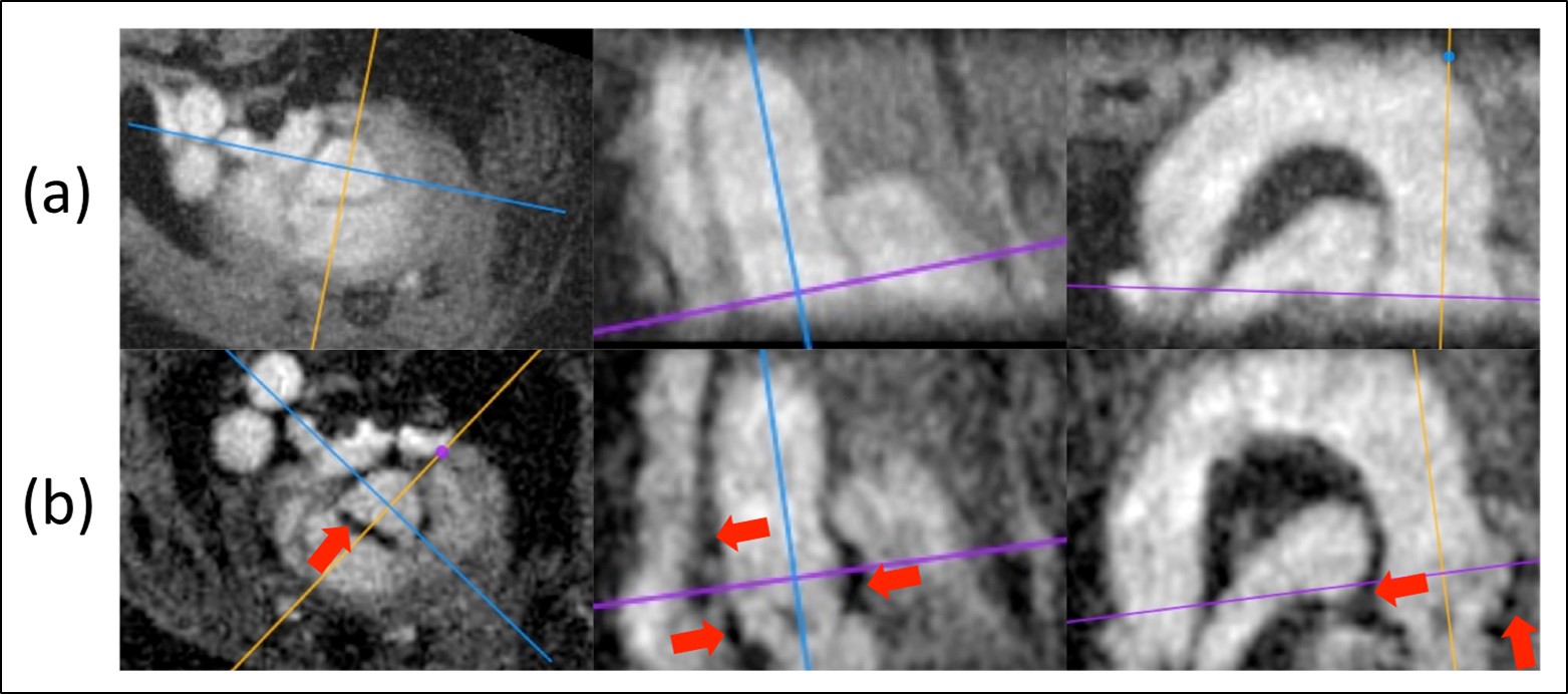
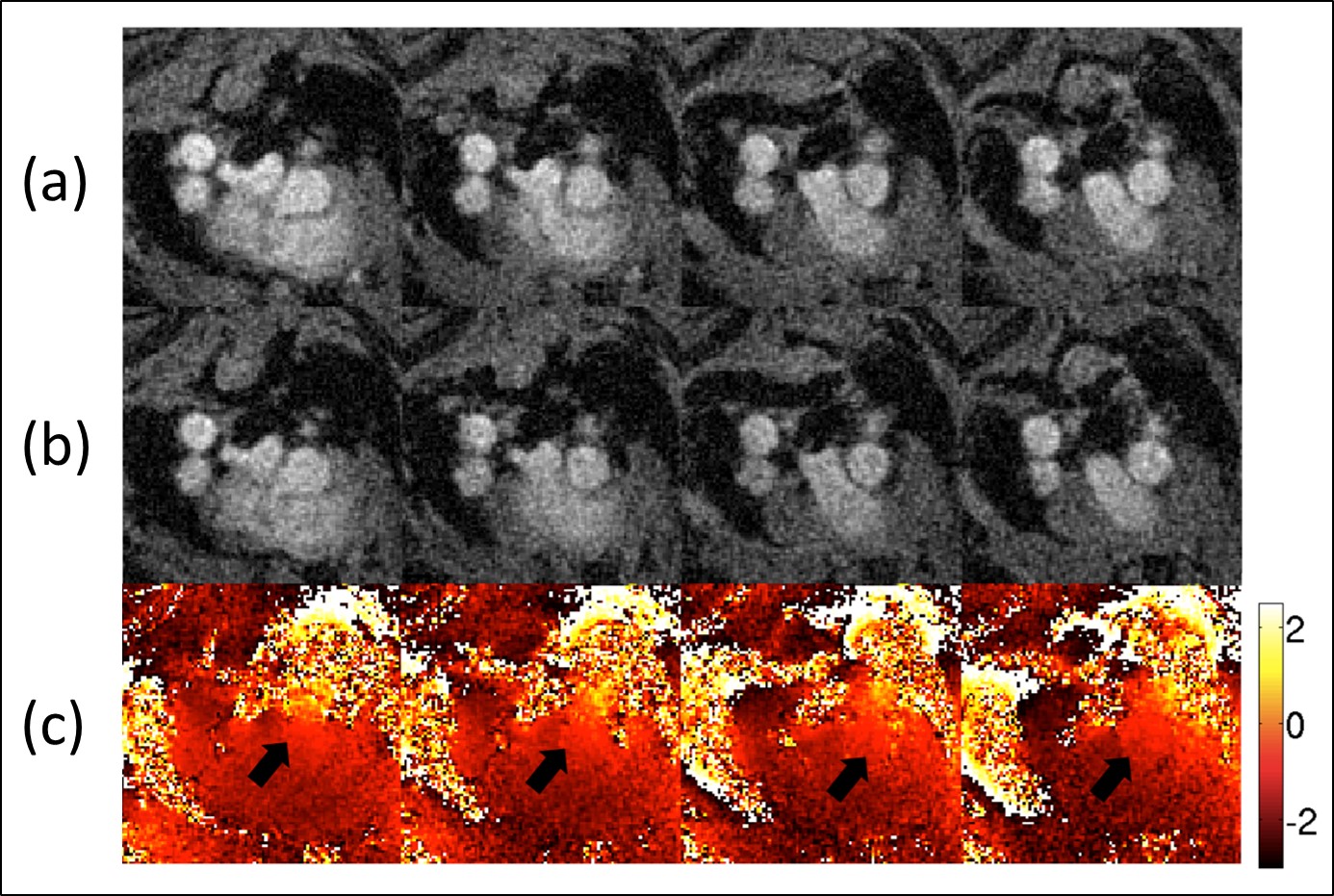
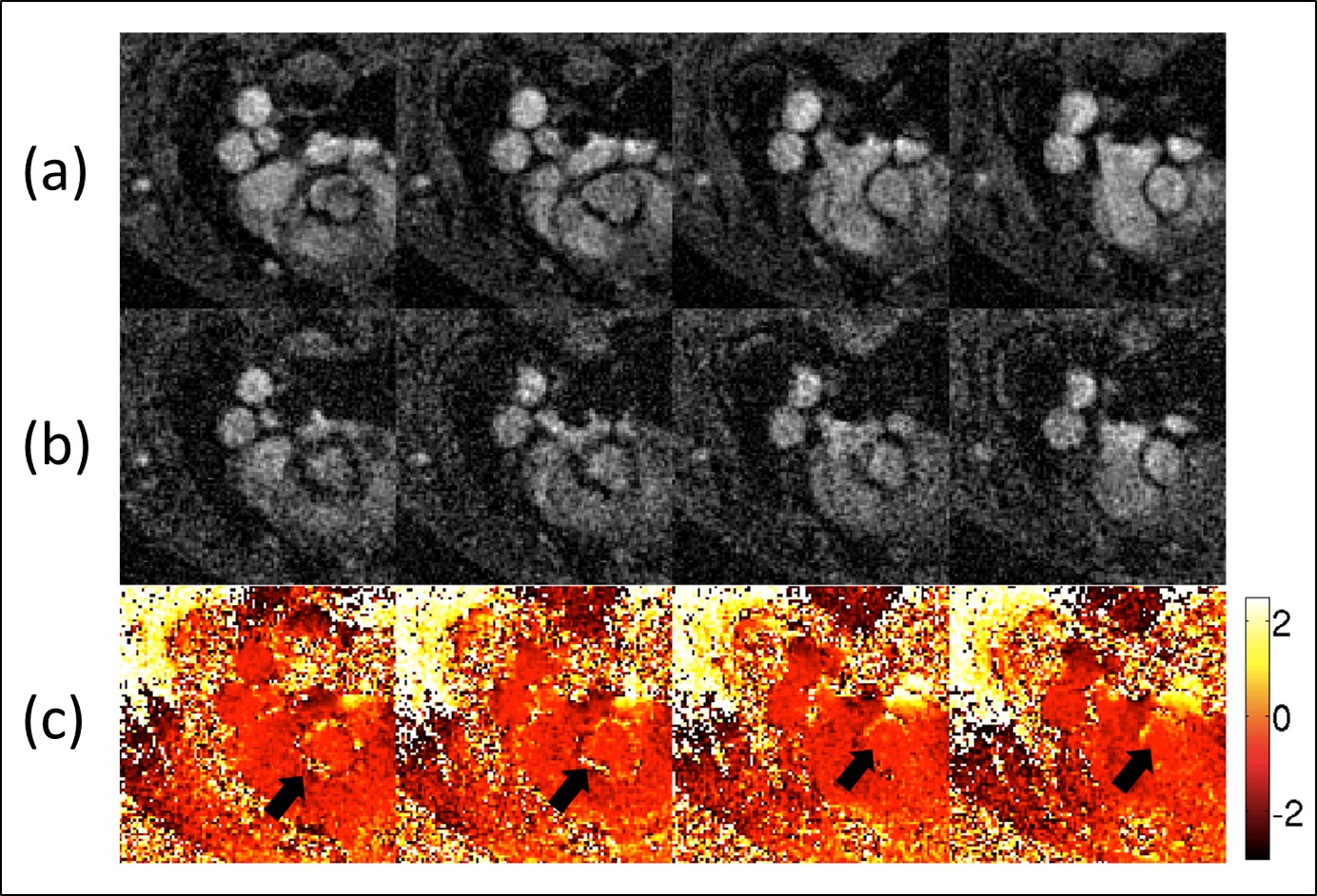
Fig.1 illustrates three slice orientations of the retrospective radial 3D-Cine measurement (TE=1.5 ms) before (top) and after (bottom) administration of the contrast agent. The post-CA measurement indicates significant signal losses in the aortic root and the ascending aorta due to the presence of the iron particles. The longitudinal views of the aortic arch show that these signal cancelations can be found mostly in the aortic root and along the ascending aorta. Fig. 2a&b shows 4 slices along the ascending aorta obtained from the baseline measurement at 2 different echo times. In the phase difference map (Fig. 2c), no significant local phase differences could be found in the aortic vessel wall. Fig. 3 displays the results of the post-CA measurement. The phase difference map (Fig 3c) shows significant phase jumps all along the ascending aorta, which can be referred to the local susceptibility differences.Discussion and Conclusion
In this abstract we demonstrate that the coverage of the complete aortic arch at high spatial resolution and the detection of functionalized USPIOs is feasible with an ECG-free radial 3D-Cine acquisition. The measurement time was only 12.5 minutes for one 3D dataset, which is two times the measurement needed for the standard single-slice 2D-protocol1. The cine reconstruction enables the tracking of the nanoparticles during the complete cardiac cycle and the measurement of two different gradient echoes provides the calculation of phase difference maps. Both are valuable supplementary information to differentiate signal losses caused by iron particles from other signal cancelations (e.g. caused by blood flow or lung tissue). Furthermore, this method provides the possibility to correlate the localization of inflammation sites with measurements of endothelial wall shear stress in order to study its potential causal relations.Acknowledgements
This work was supported by grants from the Deutsche Forschungsgemeinschaft (SFB 688 B5, Z2), the Bundesministerium für Bildung und Forschung (BMBF01 EO1004) and the Comprehensive Heart Failure Center (CHFC)References
1. Michalska et al., Arteriosc Thromb Vasc Biol 32(10): 2350-2357.
2. Winter et al., JCMR, [2013], 15:88-98
3. Winter et al., Magn Reson Med, [2016]; DOI: 10.1002/mrm.26068
Figures