2764
Dual identity of the interventricular septum with in vivo diffusion tensor imaging1Cardiovascular BRU, Royal Brompton Hospital, London, United Kingdom, 2NHLBI, National Institutes of Health, MD, United States
Synopsis
Heart muscle has a complex cellular helical arrangement, where bundles of myocytes, known as sheetlets interleave with collagen-lined shear layers. The right and left ventricle form separately in early stages of cardiogenesis, resulting in a dual inter-ventricular septum. In this work we compare the myocyte and sheetlet microstructure in the septum and LV free-wall in healthy and HCM hearts with in vivo diffusion tensor imaging. Results show that the right-side of the interventricular-septum has myocyte orientations not seen in the free-wall, and also lower sheetlet angles, which may indicate an RV identity in this region.
Purpose
Compare the myocyte and sheetlet microstructure in the interventricular septum and LV free-wall in healthy and HCM hearts with in vivo diffusion tensor imaging (DTI).Background
The micro-structure of the developed heart muscle is complex. Myocytes exhibit a helical organization with helix angles rotating smoothly across the heart wall 1 2. Additionally, myocytes have a laminar organization, comprising of sheetlets, which are bundles of myocytes, and collagen lined fissures 3 4 5.
The right and left ventricle form separately in early stages of cardiogenesis, resulting in a dual inter-ventricular septum (IVS)6. In the fully formed human heart, the two ventricles are coupled together and its shape and curvature are defined by biventricular pressure and volume.
In this work we compare the myocyte and sheetlet microstructure in the IVS and LV free-wall in healthy and HCM hearts with in vivo diffusion tensor imaging (DTI).
Methods
DTI data of a mid-ventricular slice from 19 healthy and 13 HCM hearts scanned at 3T (Siemens Healthcare, Erlangen, Germany) during the systolic pause was used. A STEAM sequence: 6 diffusion directions at b = 600 and 150 s/mm2, fat saturation, TR = 2RR-intervals, TE = 23 ms, SENSE (2 x acceleration), echo train duration 13 ms, reconstructed spatial resolution = 1.4 × 1.4 mm2, 10–15 averages.
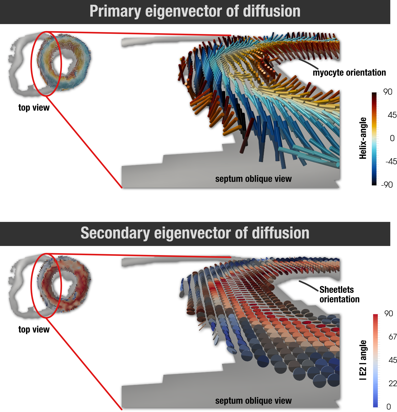
The diffusion tensor orientation of the primary and secondary eigenvectors was calculated. These parameters have been shown to respectively approximate the orientation of myocyte long-axis known as helix-angle (HA), and sheetlets defined here as E2A.
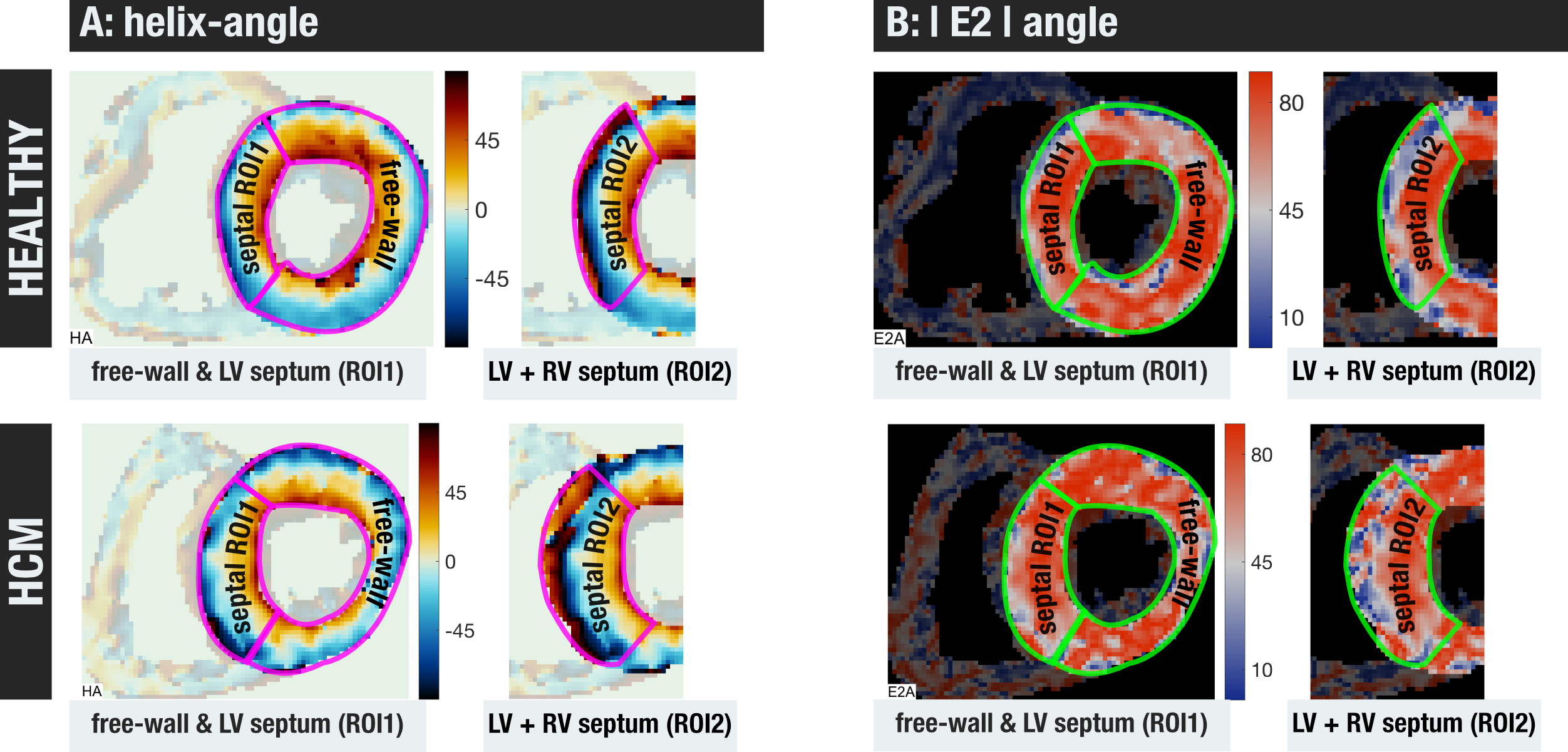
Three left ventricular regions were delineated based on the HA image: the free-wall, distal to the RV; the septal segment containing only the same myocyte orientations as the free-wall, considered to be the LV septum (ROI1); and finally, the entire interventricular septal wall (ROI2) (figure 1).
Results
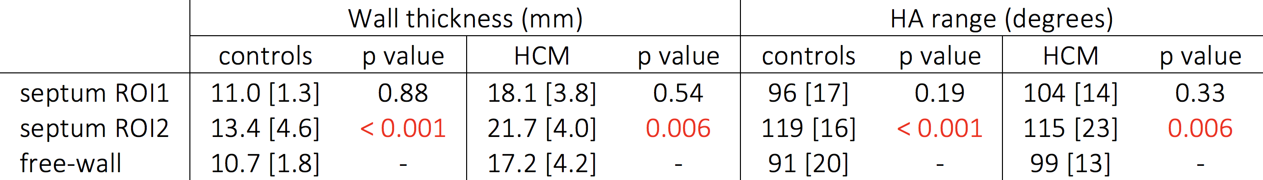
The LV septum comprises approximately 80% of the total IVS thickness. The range of helix angles of the entire IVS wall (ROI2) is significantly higher than the free-wall (figure 2). Both the free-wall and entire septum show smooth transmural transition of myocyte orientations, although with the higher range of helix-angles in the IVS, the myocytes wrap back to positive angles on the RV side, not seen on the epicardium of the free wall (figure 3).
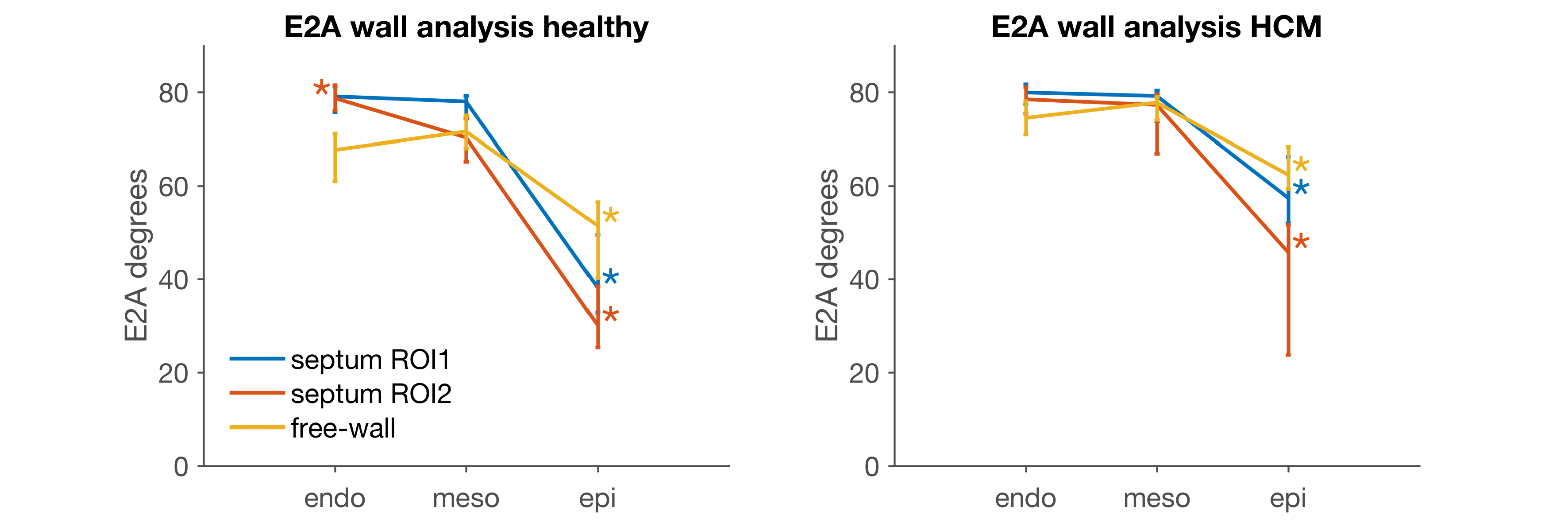
Additionally, there are consistently lower E2As for the epicardial layer, especially for the RV side when considering the entire IVS (figure 3 & 4).
Discussion & conclusions
DTI has shown that the right-side of the IVS has myocyte orientations not seen in the free-wall, and lower E2As. There was no obvious interventricular border from the DTI data, although these microstructural differences may indicate an RV identity to this region.
Cardiac MR analysis quite often involves the delineation of the LV. Even though there is no clear delineation of the septal LV, even microstructurally, the dual identity of the IVS must be considered, as there are clear implications for many different MR derived parameters. For example, maximal wall thickness measurements for the diagnosis of HCM and myocardial first-pass perfusion quantification.
Acknowledgements
This project was supported by the NIHR cardiovascular Biomedical Research Unit at the Royal Brompton and Harefield NHS Foundation Trust.References
1 – Pettigrew, J. B. On the Arrangement of the Muscular Fibres in the Ventricles of the Vertebrate Heart, with Physiological Remarks. Philosophical Transactions of the Royal Society of London (1864): 445-500.
2 – Streeter, Jr, D. D., Spotnitz, H. M., Patel, D. P., et al. Fiber orientation in the canine left ventricle during diastole and systole. Circ Res (1969): 339-47.
3 – Harrington, K. B., Rodriguez, F., Cheng, A., et al. Direct measurement of transmural laminar architecture in the anterolateral wall of the ovine left ventricle: new implications for wall thickening mechanics. Am J Physiol Heart Circ Physiol (2005): H1324-30.
4 – Gilbert, S. H., Benoist, D., Benson, A. P., et al. Visualization and quantification of whole rat heart laminar structure using high-spatial resolution contrast-enhanced MRI. Am J Physiol Heart Circ Physiol (2012): H287-98.
5 – Hales, P. W., Schneider, J. E., Burton, R. A. B., et al. Histo-anatomical structure of the living isolated rat heart in two contraction states assessed by diffusion tensor MRI. Prog Biophys Mol Biol (2012): 319-30.
6 - Franco, D., Meilhac, S. M., Christoffels, V. M., et al. Left and right ventricular contributions to the formation of the interventricular septum in the mouse heart. Dev Biol (2006): 366-75.
Figures