2753
A Versatile MOLLI-based Inversion Time (TI) scout sequence for Myocardial Nulling Determination in emerging Late Gadolinium Enhanced CMR Variants: A Phantom Study1Medicine, The University of Chicago, Chicago, IL, United States, 2Radiology, The University of Chicago, Chicago, IL, United States, 3Philips HealthTech, Cleveland, OH, United States
Synopsis
A Look-Locker scout is used for optimal inversion time (TI) determination of myocardial nulling in Late Gadolinium Enhancement (LGE) Cardiac Magnetic Resonance. Recently, novel LGE technique variants such as the Wideband approach have been proposed, and these approaches can lead to variability in the signal evolution between the Look-Locker TI-scout and LGE. In this study, we propose a Modified Look-Locker Inversion (MOLLI)-based TI-scout that closely matches its signal evolution to that of any LGE variant. Bloch simulation and phantom evaluation are performed to compare the null-times from the conventional LL-based and proposed MOLLI-based approaches against the LGE protocol over a range of clinically relevant T1 values observed in viability imaging.
Introduction
The Look-Locker (LL) scout that employs either a low-flip-angle Spoiled-Gradient-Echo (SPGR) or a balanced Steady-State-Free-Precession (b-SSFP) readout is the preferred method for optimal Inversion Time (TI) determination for myocardial nulling in Late Gadolinium Enhancement (LGE) CMR (Ref1). Recently, new LGE variants, such as the Wideband (WB) approach for overcoming scar-mimicking, hyperintense artifacts from implantable cardiac defibrillators (ICD) (Refs2-4) have become more prevalent. As new LGE variants enable CMR viability assessments in patients with ICDs, the complexity of the viability CMR protocol, including the proper null-time determination using TI-scouts also increases. For example, b-SSFP-based scouts become infeasible, and SPGR-based LL-scouts must be properly examined in terms of its ability to match the observed nulling in the LGE variant across a range of clinically relevant T1s at different heart-rates, flip-angles, and Inversion RF pulse efficiency values (Ref3).
In this study, we propose a simple, and potentially versatile alternative to the conventional SPGR-LL method for TI-scout imaging. Based on Modified Look-Locker Inversion (MOLLI) (Ref6), our approach replicates the longitudinal signal evolution as the LGE variant during TI-scout. We first evaluate the proposed approach using Bloch simulation, and next examine the proposed MOLLI-based TI-scout in a phantom using the Wideband-LGE protocol.
Methods
Proposed sequence: We propose a 2RR-cyclic MOLLI TI-scout sequence using matched Inversion RF and SPGR readout as the LGE, analogous to [1]-(1)-[1]-(1)-…-(1)-[1] SPGR-MOLLI scheme. The [1-acq]-(1-rest) 2RR-cycle is performed over (2N-1) heartbeats, yielding N scout steps.
Simulation: An exhaustive Bloch simulation (Ref7) that varied the inversion-efficiency [0.8-1.0], TI [150-500ms], and RR-interval [600-1500ms] was first performed to characterize signal evolution. The null-points at various T1 ranges [100-1000ms] were compared between the conventional LL-based TI-scout, the proposed approach, and the LGE reference. Additionally, the extent of signal changes due to TR/#phase-encodes differences between the SPGR-MOLLI-scout and LGE was examined.
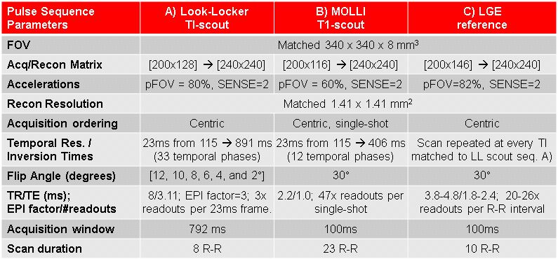
Phantom study: Imaging was performed at both 1.5T and 3T (Philips Achieva) using 5 and 32-channel cardiac arrays, respectively. The phantom contained 28 vials at different Gd and agarose concentrations. Three sequences were performed using Wideband IR: A) the WB-LL TI-scout, B) proposed 2RR WB-MOLLI TI-scout, and C) WB-LGE repeated with TIs matched to each scout step. Acquisition parameters are outlined in Table 1; B) and C) were matched as closely as possible for similar signal evolution. Regions-of-interest for each vial was delineated for null-time analysis in A)-C).
Results
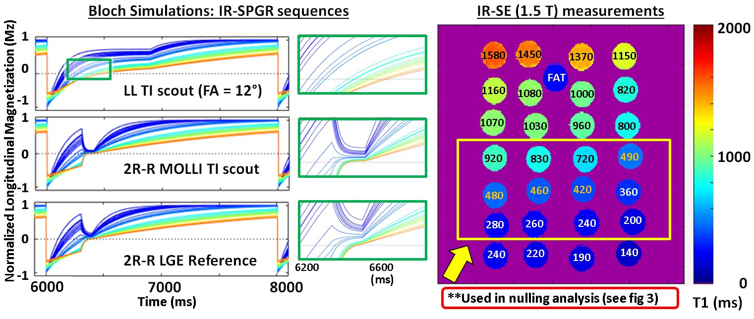
Figure 1 shows a representative Bloch simulation of the three IR-SPGR sequences. Here, the TI values for B) and C) is matched at 305ms. The LL-approach at FA=12° underestimated the optimal null-times for target vials at T1 = 460-490ms, whereas the LGE reference and MOLLI-based TI-scout schemes both yielded matched null-points (Nulled TI: LL = 230ms vs LGE/MOLLI = 305ms in this figure).
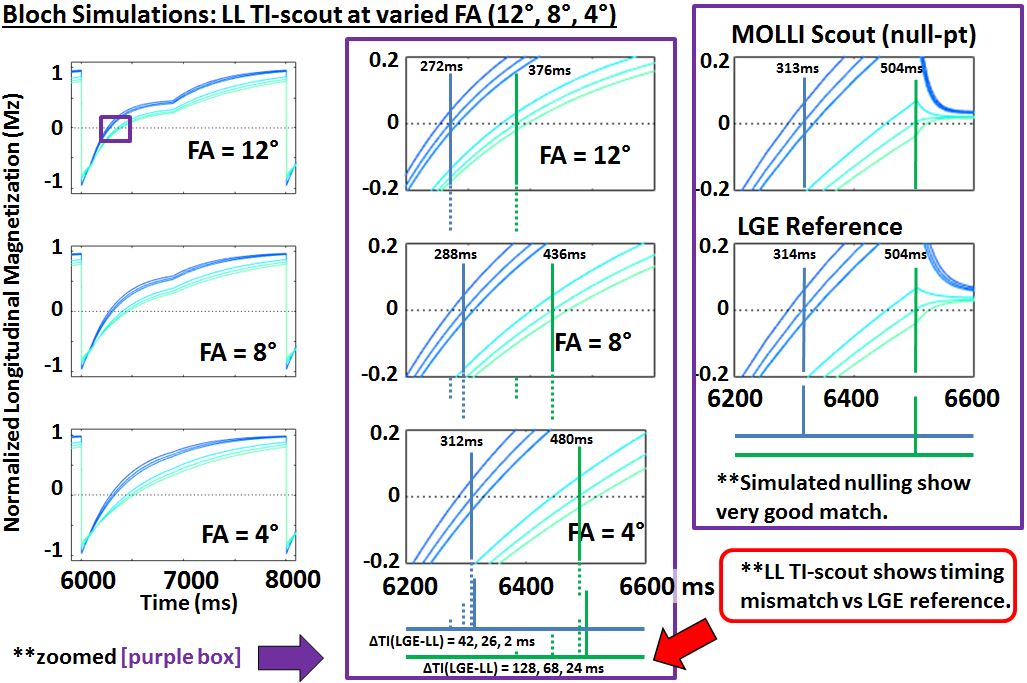
Figure 2 shows the LL-based TI-scout using three different flip angles (FA = 12°, 8°, 4°), where a lower flip-angle yielded improved TI agreement with simulated myocardial nulling exhibited in LGE; this concurred with Ref4. Upon an exhaustive simulation, the maximal normalized Mz difference between the LGE variant and MOLLI TI-scout at the initial readout for vials with T1=[400-900ms] was 0.67% across over 35000 parameter combinations; showing excellent agreement despite the difference in TR/#phase-encodes, which was 3.7ms/26 for LGE, and 2.1ms/50 for SPGR-MOLLI TI-scout.
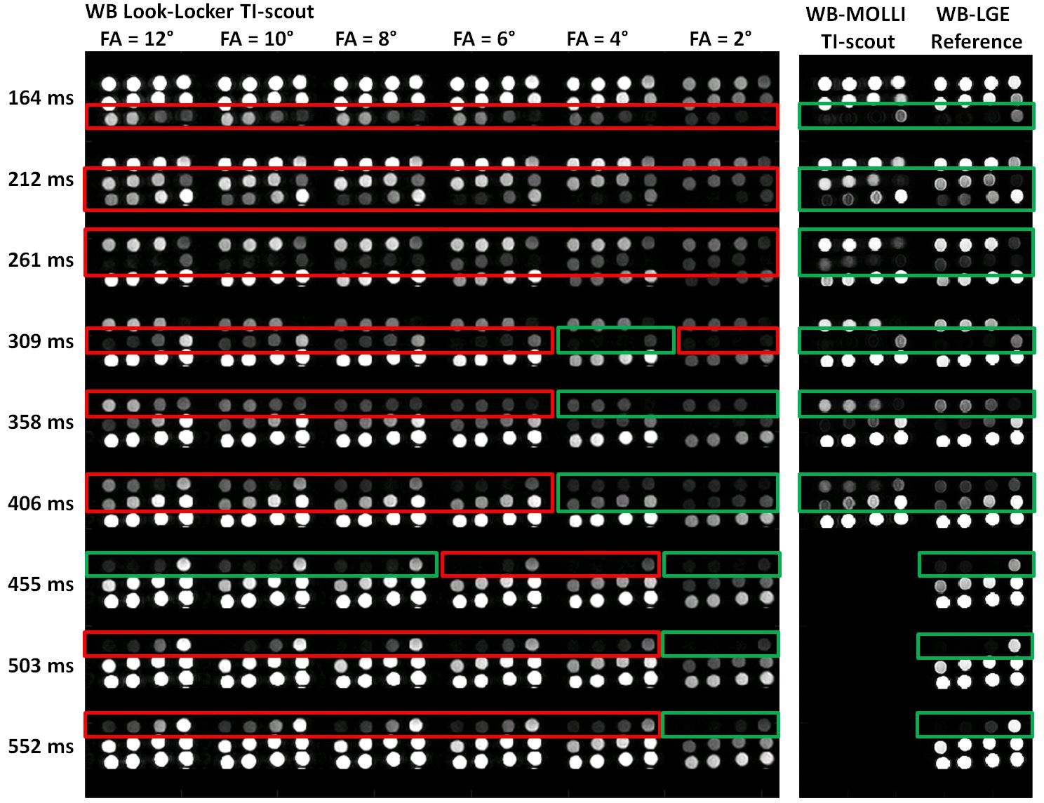
Figure 3 shows the phantom images acquired with a 3.8kHz bandwidth Wideband-IR scheme on the 3T system. The retrospectively computed inversion-efficiency was delta=(0.83/0.81) at (1.5/3.0T), respectively, using customized set of WB-RF beta/μ parameters (Ref2). With this potentially reduced inversion pulse for an ideal LL-assumption of delta=1 (Ref7), linear regression was performed on n=9 vial subset in which the reference LGE-derived nulling was observed between [150-400ms]. All WB-LL TI-scouts at flip-angles between 2°-12° underestimated the WB-LGE-derived optimal TI by 5-35%; nulling for each vial was underestimated by 23, 48, or 70ms at the acquired temporal resolution. On the other hand, the WB-MOLLI TI-scout correctly derived the LGE-based nulling points for each vial to within the TI step. The derived TI differences versus the WB-LGE reference was statistically significant (P<0.0005; N=17 total vials; 9@1.5T, 8@3.0T).
Discussion
The feasibility of the proposed 2RR-cyclic SPGR-MOLLI TI-scout in capturing the optimal nulling times is shown using an exhaustive Bloch simulation and using an inversion-inefficient WB-LGE protocol on a phantom. This MOLLI-based TI-scout replicates the tissue signal evolution in LGE more accurately than conventional LL-based TI-scouts, and thus provides more consistent nulling times as the LGE reference. Finally, this scout approach may help loosen strict acquisition conditions as new LGE variants are introduced; including the problem of IR and readout RF designs for these new Wideband protocols.
Acknowledgements
This work was supported by the NCATS Institute of Translational Medicine Pilot Award (PI. K. Kawaji CTSA UL1 TR000430-9).References
1. Simonnetti et al. Radiology 2001.
2. Rashid et al. Radiology 2014.
3. Rashid et al. Proc. ISMRM 2016. pp3169.
4. Rashid et al. JCMR 2014. 16(S1):P019.
5. Ogawa et al. J of Japanese Radiographic Soc. 2011.
6. Messroghli et al. MRM 2004.
7. Deichmann et al. JMR 1992.
Figures