2748
Myocardial fat quantification of normal subjects via 7-peaks mDixon model1Executive Health Management Center, Cheng Ching Hospital, Taichung, Taiwan, 2Philips Healthcare, Taipei, Taiwan, 3Philips Healthcare, Hong Kong
Synopsis
Cardiac magnetic resonance imaging provides a lot of physiological information about myocardial pathology, and the mDixon technique also has been developed on cardiac application recently. The fat deposition in myocardium is possibly related with cardiomyopathy, so it is important to determine the fat composition of myocardium. The mDixon provides an easy way to acquire such this information within one breath hold, and it has a consistent result with magnetic resonance spectroscopy from the previous study. Also it has been reported that multipeak fat spectrum model gives more robustness, and 7-peaks model is used in this study. The aim of this study is to evaluate fat fraction of myocardium with 7-peaks model.
Purpose
Cardiac magnetic resonance imaging provides a lot of physiological information about myocardial pathology, and the mDixon technique also has been developed on cardiac application1 recently. The fat deposition in myocardium is possibly related with cardiomyopathy, so it is important to determine the fat composition of myocardium. The mDixon provides an easy way to acquire such this information within one breath hold, and it has a consistent result with magnetic resonance spectroscopy from the previous study2. Also it has been reported that multipeak fat spectrum model gives more robustness3, and 7-peaks model is used in this study. The aim of this study is to evaluate fat fraction of myocardium with 7-peaks model.Material and method
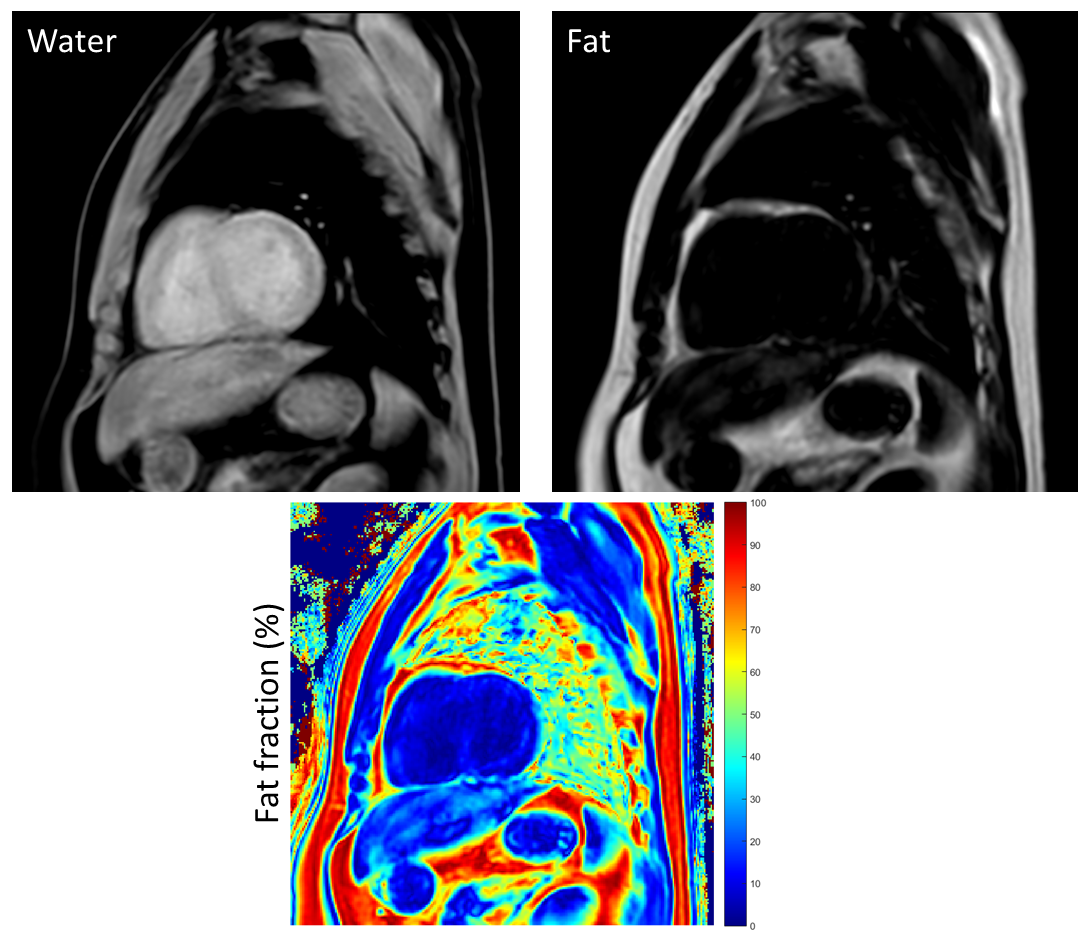
10 healthy subjects (average 40.6 years old, 24~7, 5 females and 5males) participated in this study, after signing the inform consent. All subjects were examined on Philips Ingenia 1.5T MR system (Philips Healthcare, Eindhoven, The Netherlands) with a 32 channel torso coil. 3D mDixon cardiac imaging uses dual-echo turbo field echo with ECG triggered, and parameters are TR/TE = 5.1/[1.4 3.0]ms, flip angle = 10 degrees, field of view = 30 cm, slice thickness = 10 mm, slice number = 10, SENSE factor=2X2. Water and fat images were calculated with 7-peaks model, exported directly from the console. The fat fraction (FF) map is calculated with the equation : FF = fat/(water + fat). ROIs were selected at the septal area, and the fat fraction was calculated within ROIs. FF map and ROIs was processed with home-made script and GUI on MATLAB 2015a (The Mathwork, Natick, MA) platform.Result
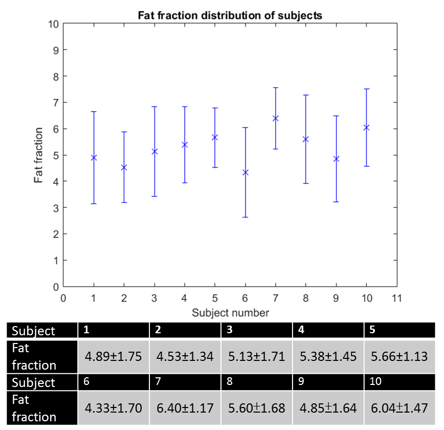
Acquired images of cardiac mDixon are shown in figure 1, including water, fat image and FF map. The averaged FF of all subjects is 5.28±0.66%, and FFs of all subjects are described in figure 2. All FFs show the similar distribution.Discussion and conclusion
It shows a stable distribution among all subjects from figure 2. Fat fractions of normal subjects reported from previous studies vary from 0.1~5%245, and our results are slightly higher than others. This difference is possibly caused by different fat and water spectral model or the effect of T1 or T2 correction. 7-peaks model used in this study gives a better separation of fat and water than single peak model3, and therefore this might be the reason yielding a higher level of the fat fraction. In conclusion, 7-peaks model mDixon imaging demonstrates a stable result of fat quantification within one breath hold among all subjects, and it still needs more subjects to validate and myocardial deposition cases to test in the future.Acknowledgements
No acknowledgement found.References
1. Kellman, Peter, et al. "Multiecho dixon fat and water separation method for detecting fibrofatty infiltration in the myocardium." Magnetic resonance in medicine 61.1 (2009): 215-221.
2. Liu, Chia-Ying, et al. "Myocardial fat quantification in humans: evaluation by two-point water-fat imaging and localized proton spectroscopy." Magnetic resonance in medicine 63.4 (2010): 892-901.
3. Kijowski, Richard, et al. "Improved fat suppression using multipeak reconstruction for IDEAL chemical shift fat-water separation: Application with fast spin echo imaging." Journal of Magnetic Resonance Imaging 29.2 (2009): 436-442.
4. Venkatesh, Bharath Ambale, et al. "MR proton spectroscopy for myocardial lipid deposition quantification: a quantitative comparison between 1.5 T and 3T." Journal of Magnetic Resonance Imaging 36.5 (2012): 1222-1230.
5. Venkatesh, Bharath Ambale, et al. "MR proton spectroscopy for myocardial lipid deposition quantification: a quantitative comparison between 1.5 T and 3T." Journal of Magnetic Resonance Imaging 36.5 (2012): 1222-1230.
Figures