2719
Simultaneous measurements of myocardium T1, T2 map, Cine and synthetic LGE using Inversion Recovery tiny golden angle radial balanced-SSFP within 6 secondPanki Kim1 and Byoung Wook Choi1
1Department of Radiology, Severance Hospital, Yonsei University College of Medicine, Seoul, Korea, Republic of
Synopsis
In the cardiac MRI study, the quantification of T1, T2 relaxation time has become an important indication as well as the cine and the late gadolinium enhancement. In this work, we presented a novel method for simultaneous acquisition of T1, T2 map and Cine image of the myocardium based on the transient phase of inversion recovery balanced-SSFP imaging, and was proven potential both phantom and in vivo on a healthy volunteer.
Introduction
In the cardiac MRI study, the quantification of T1, T2 relaxation time has become an important indication as well as the cine and the late gadolinium enhancement. However, each technique requires definitely a breath-holding and breathing space to freeze respiratory motion. This waiting time might extend an overall scan time and make patients uncomfortable. To reduce the total scan time, we proposed a novel sequence to simultaneously measure the T1, T2 relaxation time, spin density, cine and late-gadolinium enhancement imaging(LGE) using a tiny golden angle radial balanced-SSFP with inversion recovery pulse (IR-tGAR-bSSFP) within 6 second.Methods
The inversion recovery balanced-SSFP can simultaneously quantify both T1 and T2 relaxation time as previous studies1. The Inversion recovery bSSFP analytical formulation can be described as follows; $$S(t)=S_{stst}(1-(S_0+S_{stst})\cdot exp(-\frac{t}{T_1^*}))$$ , where Sstst is the steady-state signal; S0 is the transient state signal; and T1* is the apparent relaxation time; T1, T2, and proton density(M0) can be obtained from a pixelwise nonlinear fitting as accordings; $$T_1=T_1^*\frac{S_0}{S_{stst}}\cos(\frac{\alpha}{2})$$, $$T_2=T_1^*\sin^{2}(\frac{\alpha}{2})(1-\frac{S_{stst}}{S_0}\cos(\frac{\alpha}{2}))^{-1}$$, $$M_0=\frac{S_0}{\sin(\frac{\alpha}{2})}$$where the excitation flip angle . In order to achieve the simultaneous T1, T2 and Cine imaging, we developed a tiny golden-angle (23.62°) radial balanced-SSFP with inversion recovery(IR) pulse to reduce strong image artefacts due to eddy currents caused by the large angular increment of the golden-angle (111.25°) on a 3.0T MRI (Siemens, Tim Trio, Germany) as illustrated in Figure 1. To give heavy IR-weighting on diastolic phase, the IR pulse was just before the diastolic phase by the trigger-delay, subsequently, the tiny golden angle radial balanced-SSFP imaging performed without ECG triggering. For image-based cardiac self-gating, low-spatial and high-temporal resolution sliding-window images were reconstructed by a nonuniform fast Fourier transform (NUFFT). Cine images were reconstructed by compress-sensing algorithm3 over last two cardiac phases based on end-diastolic phases. To calculate the T1 map, the IR-weighted images on each diastolic phase were reconstructed by k-space weighted image contrast (KWIC) filtering method with highly-temporal resolution, and then a motion correction algorithm4 was applied to minimize physiological motion and to improve the accuracy of T1 estimation. The IR-tGAR-bSSFP was performed as following parameters: TR/TE=2.57/1.36ms, flip angle = 35°, matrix = 128x128, field-of-view = 250x250 mm2, slice thickness = 8mm, receiver bandwidth = 1502 Hz/pixel, total number of projections = 2300, total experiment time = 5.9 sec. The sequence was validated both T1, T2 phantom and a healthy volunteer by comparing it with the Modified Look-Locker Inversion Recovery(MOLLI) with 5-3-3 pattern, the multiple T2-prepared-bSSFP and a conventional retrospectively gated bSSFP Cine image. T1 and T2 phantom compartments contained Dotarem® (Guerbet, France) solutions and agarose at different amounts yielding a range of T1 and T2 values similar to in vivo. T1 and T2 values were assessed through Bland-Altman plots were drawn to show the agreement between MOLLI, T2-prepared bSSFP and the IR-tGAR-bSSFP methods.Results
In Fig. 2, the measured range of T1 and T2 values in phantoms was 97-2200 ms and 29-270 ms, respectively. The Bland-Altman plots showed a bias 6.03 ms for T1 with 95% limits of agreement ±24.58ms, and a bias of 5.87 ms for T2 with 95% limits of agreement ±33.41 ms. In Fig.3, T1 value of myocardium and blood was 1208.9±89.2 ms and 1885.7±20.5 ms on MOLLI, and 1346.7±62.9 ms and 1768.4±28.1 ms on IR-tGAR-bSSFP, respectively. T2 value of myocardium was 45.3±8.2 ms on T2-prepared bSSFP and 46.8±17ms on IR-tGAR-bSSFP. In Fig.4, both cine images on end-systolic and diastolic phase had shown a similar image quality.Discussion and conclusions
In this work, we presented a novel method for simultaneous acquisition of T1, T2 map and Cine image of the myocardium based on the transient phase of inversion recovery balanced-SSFP imaging, and was proven potential both phantom and in vivo on a healthy volunteer. Also, in case of need, the proton density and a synthetic IR image at theoretical inversion time can be reproduced simply according to reference 5. This proposed method can produce inherently coregistered T1, T2, and, potentially, proton density maps and synthetic LGE of the myocardium within 6 second as well as Cine image.Acknowledgements
This work was supported by the National Research Foundation of Korea (RF) grant funded by the Korea government (MISP) (No. 2016R1C1B1013837)References
[1] Schmitt, Peter, et al. "Inversion recovery TrueFISP: quantification of T1, T2, and spin density." Magnetic resonance in medicine 51.4 (2004): 661-667. [2] Wundrak, Stefan, et al. "A small surrogate for the golden angle in time-resolved radial MRI based on generalized fibonacci sequences." IEEE transactions on medical imaging 34.6 (2015): 1262-1269. [3] Block, Kai Tobias, Martin Uecker, and Jens Frahm. "Undersampled radial MRI with multiple coils. Iterative image reconstruction using a total variation constraint." Magnetic resonance in medicine 57.6 (2007): 1086-1098. [4] Xue, Hui, et al. "Motion correction for myocardial T1 mapping using image registration with synthetic image estimation." Magnetic resonance in medicine 67.6 (2012): 1644-1655. [5] Varga-Szemes, Akos, et al. "Myocardial Late Gadolinium Enhancement: Accuracy of T1 Mapping–based Synthetic Inversion-Recovery Imaging." Radiology 278.2 (2015): 374-382.Figures
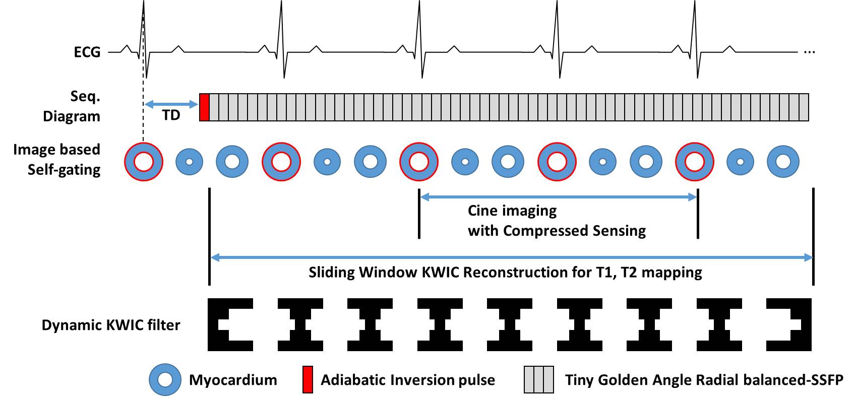
Scheme of the
inversion-recovery tiny Golden-Angle Radial Balanced-SSFP (IR-tGAR-bSSFP). A
single-shot acquisition is initiated in the diastolic phase after an inversion
pulse.
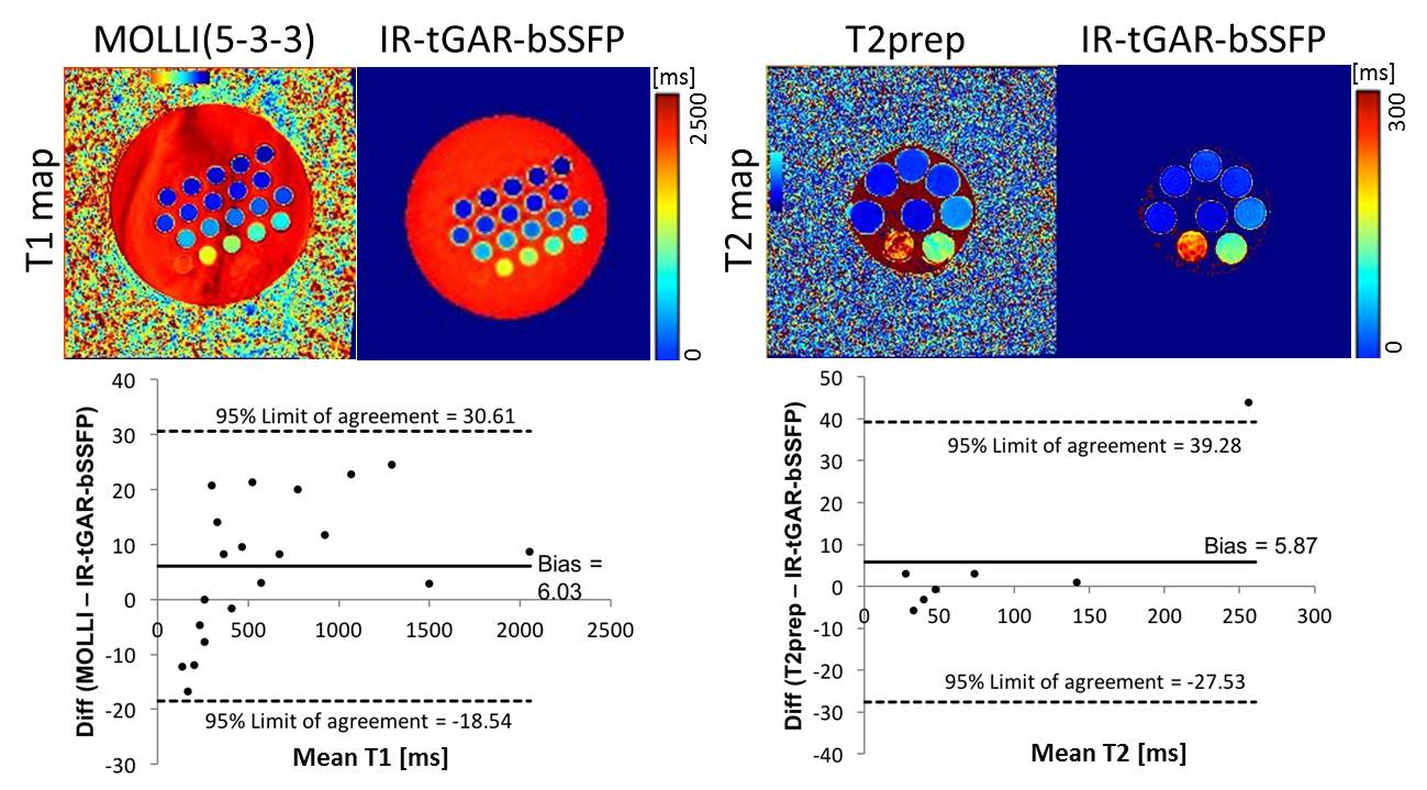
T1 and T2 maps in T1 and T2 phantom,
compared with a MOLLI T1 map and a T2-prep bSSFP T2
map, and Bland-Altman plots of the agreement.
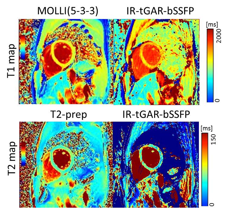
T1 and T2 map in a mid-ventricular short
axis view of a healthy volunteer, compared with MOLLI T1 map and T2-prep bSSFP T2
map.
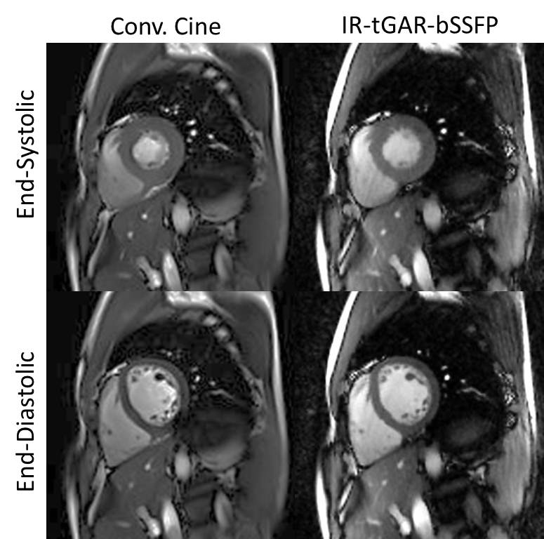
End-systolic and end-diastolic cine
images in a mid-ventricular short axis view of a healthy volunteer, compared
with a conventional retrospective ECG gated bSSFP cine
image.